 Movie
Movie Controller

[English] 日本語
 Yorodumi
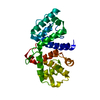
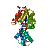
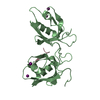
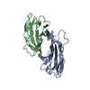
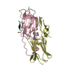
- PDB-4rey: Crystal Structure of the GRASP65-GM130 C-terminal peptide complex -
+ Open data

- Basic information
| Entry | Database: PDB / ID: 4rey | ||||||
|---|---|---|---|---|---|---|---|
| Title | Crystal Structure of the GRASP65-GM130 C-terminal peptide complex | ||||||
 Components Components |
| ||||||
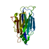
 Keywords Keywords |  MEMBRANE PROTEIN / PDZ fold six-stranded anti parallel-barrel capped by two-helices / protein interaction MEMBRANE PROTEIN / PDZ fold six-stranded anti parallel-barrel capped by two-helices / protein interaction | ||||||
| Function / homology |  Function and homology informationmeiotic spindle assembly / Golgi cis cisterna / positive regulation of protein glycosylation / Golgi disassembly / establishment of protein localization to plasma membrane / asymmetric cell division / negative regulation of dendrite morphogenesis / Golgi ribbon formation / microtubule nucleation / importin-alpha family protein binding ...meiotic spindle assembly / Golgi cis cisterna / positive regulation of protein glycosylation / Golgi disassembly / establishment of protein localization to plasma membrane / asymmetric cell division / negative regulation of dendrite morphogenesis / Golgi ribbon formation / microtubule nucleation / importin-alpha family protein binding / cis-Golgi network / protein N-linked glycosylation / Golgi Cisternae Pericentriolar Stack Reorganization / centrosome cycle / COPII-mediated vesicle transport / syntaxin binding / Golgi cisterna membrane / Deregulated CDK5 triggers multiple neurodegenerative pathways in Alzheimer's disease models / COPII-coated ER to Golgi transport vesicle / Golgi organization / protein glycosylation / mitotic spindle assembly / spindle assembly / endoplasmic reticulum to Golgi vesicle-mediated transport / COPI-mediated anterograde transport / endoplasmic reticulum-Golgi intermediate compartment membrane / negative regulation of autophagy / negative regulation of protein binding / mitotic spindle / spindle pole / protein transport / microtubule binding / protein homotetramerization / microtubule / cadherin binding / Golgi membrane / protein kinase binding / Golgi apparatus / identical protein binding / metal ion binding Function and homology informationmeiotic spindle assembly / Golgi cis cisterna / positive regulation of protein glycosylation / Golgi disassembly / establishment of protein localization to plasma membrane / asymmetric cell division / negative regulation of dendrite morphogenesis / Golgi ribbon formation / microtubule nucleation / importin-alpha family protein binding ...meiotic spindle assembly / Golgi cis cisterna / positive regulation of protein glycosylation / Golgi disassembly / establishment of protein localization to plasma membrane / asymmetric cell division / negative regulation of dendrite morphogenesis / Golgi ribbon formation / microtubule nucleation / importin-alpha family protein binding / cis-Golgi network / protein N-linked glycosylation / Golgi Cisternae Pericentriolar Stack Reorganization / centrosome cycle / COPII-mediated vesicle transport / syntaxin binding / Golgi cisterna membrane / Deregulated CDK5 triggers multiple neurodegenerative pathways in Alzheimer's disease models / COPII-coated ER to Golgi transport vesicle / Golgi organization / protein glycosylation / mitotic spindle assembly / spindle assembly / endoplasmic reticulum to Golgi vesicle-mediated transport / COPI-mediated anterograde transport / endoplasmic reticulum-Golgi intermediate compartment membrane / negative regulation of autophagy / negative regulation of protein binding / mitotic spindle / spindle pole / protein transport / microtubule binding / protein homotetramerization / microtubule / cadherin binding / Golgi membrane / protein kinase binding / Golgi apparatus / identical protein binding / metal ion bindingSimilarity search - Function | ||||||
| Biological species |  Homo sapiens (human) Homo sapiens (human) | ||||||
| Method | X-RAY DIFFRACTION / SYNCHROTRON / MOLECULAR REPLACEMENT / Resolution: 1.96 Å | ||||||
 Authors Authors | Shi, N. / Hu, F. / Li, B. | ||||||
 Citation Citation | Journal: J.Biol.Chem. / Year: 2015 Title: Structural Basis for the Interaction between the Golgi Reassembly-stacking Protein GRASP65 and the Golgi Matrix Protein GM130. Authors: Hu, F. / Shi, X. / Li, B. / Huang, X. / Morelli, X. / Shi, N. | ||||||
| History |
|
- Structure visualization
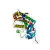
| Structure viewer | Molecule: MolmilJmol/JSmol |
|---|
- Downloads & links
-Download
| PDBx/mmCIF format | 4rey.cif.gz | 147.3 KB | Display | PDBx/mmCIF format |
|---|---|---|---|---|
| PDB format | pdb4rey.ent.gz | 119.1 KB | Display | PDB format |
| PDBx/mmJSON format | 4rey.json.gz | Tree view | PDBx/mmJSON format | |
| Others |  Other downloads Other downloads |
-Validation report
| Arichive directory | https://data.pdbj.org/pub/pdb/validation_reports/re/4reyftp://data.pdbj.org/pub/pdb/validation_reports/re/4rey | HTTPS FTP |
|---|
-Related structure data
| Related structure data |  4kfvS S: Starting model for refinement |
|---|---|
| Similar structure data |


-Links
 PDBj
PDBj


- Assembly
| Deposited unit | 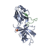
| ||||||||
|---|---|---|---|---|---|---|---|---|---|
| 1 |
| ||||||||
| Unit cell |
|
-Components
| #1: Protein | Mass: 23266.180 Da / Num. of mol.: 1 / Fragment: GRASP domain of GRAS65, UNP residues 1-210 Source method: isolated from a genetically manipulated source Source: (gene. exp.) Homo sapiens (human) / Gene: GOLPH5, GORASP1, GRASP65 / Plasmid: RSF / Production host:  Escherichia coli (E. coli) / Strain (production host): BL21(DE3) / References: UniProt: Q9BQQ3 Escherichia coli (E. coli) / Strain (production host): BL21(DE3) / References: UniProt: Q9BQQ3 |
|---|---|
| #2: Protein/peptide | Mass: 3060.392 Da / Num. of mol.: 1 / Fragment: GM130 C-TERMINAL DOMAIN, UNP residues 980-1002 Source method: isolated from a genetically manipulated source Source: (gene. exp.) Homo sapiens (human) / Gene: GM130, GOLGA2 / Plasmid: pGEX-6P-1 / Production host: Escherichia coli (E. coli) / Strain (production host): BL21(DE3) / References: UniProt: Q08379 |
| #3: Chemical | ChemComp-SO4 / Sulfate  Mass: 96.063 Da / Num. of mol.: 1 / Source method: obtained synthetically / Formula: SO4 Mass: 96.063 Da / Num. of mol.: 1 / Source method: obtained synthetically / Formula: SO4 |
| #4: Water | ChemComp-HOH / Water Mass: 18.015 Da / Num. of mol.: 201 / Source method: isolated from a natural source / Formula: H2O Mass: 18.015 Da / Num. of mol.: 201 / Source method: isolated from a natural source / Formula: H2O |
-Experimental details
-Experiment
| Experiment | Method: X-RAY DIFFRACTION / Number of used crystals: 1 |
|---|
- Sample preparation
| Crystal | Density Matthews: 3.09 Å3/Da / Density % sol: 60.21 % |
|---|---|
| Crystal grow | Temperature: 291 K / pH: 8.5 Details: 30% PEG 4000, 0.1M Tris, 0.2M Lithium sulfate, , pH 8.5, VAPOR DIFFUSION, SITTING DROP, temperature 291K |
-Data collection
| Diffraction | Mean temperature: 200 K |
|---|---|
| Diffraction source | Source: SYNCHROTRON / Site: SSRF  / Beamline: BL17U / Wavelength: 0.9791 / Beamline: BL17U / Wavelength: 0.9791 |
| Detector | Type: ADSC QUANTUM 315 / Detector: CCD / Date: Dec 9, 2013 |
| Radiation | Monochromator: SI 111 CHANNEL / Protocol: SINGLE WAVELENGTH / Monochromatic (M) / Laue (L): M / Scattering type: x-ray |
| Radiation wavelength | Wavelength: 0.9791 Å / Relative weight: 1 |
| Reflection | Resolution: 1.96→64.84 Å / Num. obs: 25936 / % possible obs: 99.56 % / Observed criterion σ(I): 1 / Redundancy: 13.1 % / Biso Wilson estimate: 32 Å2 / Rmerge(I) obs: 0.1033 / Net I/σ(I): 13.78 |
| Reflection shell | Resolution: 1.96→2.03 Å / Redundancy: 13.4 % / Rmerge(I) obs: 0.4814 / Mean I/σ(I) obs: 4.39 / % possible all: 100 |
- Processing
| Software |
| ||||||||||||||||||||||||||||||||||||||||||||||||||||||||||||||||||||||||||||||||||||||||||||||||||||||||||||||||||||||||||||||||||||||||||||||||||||||||||||||||||||||||||||||||||||||||||||||||||||||||||||||||||||||||||||||||||||||||||||||||||||||||||||||||||||||||||||||||||||||||||||||||||||||||||||||||||||||||||||||||||||||||||||||||||||||||||||||
|---|---|---|---|---|---|---|---|---|---|---|---|---|---|---|---|---|---|---|---|---|---|---|---|---|---|---|---|---|---|---|---|---|---|---|---|---|---|---|---|---|---|---|---|---|---|---|---|---|---|---|---|---|---|---|---|---|---|---|---|---|---|---|---|---|---|---|---|---|---|---|---|---|---|---|---|---|---|---|---|---|---|---|---|---|---|---|---|---|---|---|---|---|---|---|---|---|---|---|---|---|---|---|---|---|---|---|---|---|---|---|---|---|---|---|---|---|---|---|---|---|---|---|---|---|---|---|---|---|---|---|---|---|---|---|---|---|---|---|---|---|---|---|---|---|---|---|---|---|---|---|---|---|---|---|---|---|---|---|---|---|---|---|---|---|---|---|---|---|---|---|---|---|---|---|---|---|---|---|---|---|---|---|---|---|---|---|---|---|---|---|---|---|---|---|---|---|---|---|---|---|---|---|---|---|---|---|---|---|---|---|---|---|---|---|---|---|---|---|---|---|---|---|---|---|---|---|---|---|---|---|---|---|---|---|---|---|---|---|---|---|---|---|---|---|---|---|---|---|---|---|---|---|---|---|---|---|---|---|---|---|---|---|---|---|---|---|---|---|---|---|---|---|---|---|---|---|---|---|---|---|---|---|---|---|---|---|---|---|---|---|---|---|---|---|---|---|---|---|---|---|---|---|---|---|---|---|---|---|---|---|---|---|---|---|---|---|---|---|---|---|---|---|---|---|---|---|---|---|---|---|---|---|---|---|---|---|---|---|---|---|---|---|---|---|---|---|---|---|---|---|---|
| Refinement | Method to determine structure: MOLECULAR REPLACEMENT Starting model: PDB ENTRY 4KFV Resolution: 1.96→64.86 Å / SU ML: 0.17 / Phase error: 18.44 / Stereochemistry target values: ML
| ||||||||||||||||||||||||||||||||||||||||||||||||||||||||||||||||||||||||||||||||||||||||||||||||||||||||||||||||||||||||||||||||||||||||||||||||||||||||||||||||||||||||||||||||||||||||||||||||||||||||||||||||||||||||||||||||||||||||||||||||||||||||||||||||||||||||||||||||||||||||||||||||||||||||||||||||||||||||||||||||||||||||||||||||||||||||||||||
| Solvent computation | Shrinkage radii: 0.9 Å / VDW probe radii: 1.11 Å / Solvent model: FLAT BULK SOLVENT MODEL | ||||||||||||||||||||||||||||||||||||||||||||||||||||||||||||||||||||||||||||||||||||||||||||||||||||||||||||||||||||||||||||||||||||||||||||||||||||||||||||||||||||||||||||||||||||||||||||||||||||||||||||||||||||||||||||||||||||||||||||||||||||||||||||||||||||||||||||||||||||||||||||||||||||||||||||||||||||||||||||||||||||||||||||||||||||||||||||||
| Displacement parameters | Biso mean: 47 Å2 | ||||||||||||||||||||||||||||||||||||||||||||||||||||||||||||||||||||||||||||||||||||||||||||||||||||||||||||||||||||||||||||||||||||||||||||||||||||||||||||||||||||||||||||||||||||||||||||||||||||||||||||||||||||||||||||||||||||||||||||||||||||||||||||||||||||||||||||||||||||||||||||||||||||||||||||||||||||||||||||||||||||||||||||||||||||||||||||||
| Refinement step | Cycle: LAST / Resolution: 1.96→64.86 Å
| ||||||||||||||||||||||||||||||||||||||||||||||||||||||||||||||||||||||||||||||||||||||||||||||||||||||||||||||||||||||||||||||||||||||||||||||||||||||||||||||||||||||||||||||||||||||||||||||||||||||||||||||||||||||||||||||||||||||||||||||||||||||||||||||||||||||||||||||||||||||||||||||||||||||||||||||||||||||||||||||||||||||||||||||||||||||||||||||
| Refine LS restraints |
| ||||||||||||||||||||||||||||||||||||||||||||||||||||||||||||||||||||||||||||||||||||||||||||||||||||||||||||||||||||||||||||||||||||||||||||||||||||||||||||||||||||||||||||||||||||||||||||||||||||||||||||||||||||||||||||||||||||||||||||||||||||||||||||||||||||||||||||||||||||||||||||||||||||||||||||||||||||||||||||||||||||||||||||||||||||||||||||||
| LS refinement shell |
| ||||||||||||||||||||||||||||||||||||||||||||||||||||||||||||||||||||||||||||||||||||||||||||||||||||||||||||||||||||||||||||||||||||||||||||||||||||||||||||||||||||||||||||||||||||||||||||||||||||||||||||||||||||||||||||||||||||||||||||||||||||||||||||||||||||||||||||||||||||||||||||||||||||||||||||||||||||||||||||||||||||||||||||||||||||||||||||||
| Refinement TLS params. | Method: refined / Refine-ID: X-RAY DIFFRACTION
| ||||||||||||||||||||||||||||||||||||||||||||||||||||||||||||||||||||||||||||||||||||||||||||||||||||||||||||||||||||||||||||||||||||||||||||||||||||||||||||||||||||||||||||||||||||||||||||||||||||||||||||||||||||||||||||||||||||||||||||||||||||||||||||||||||||||||||||||||||||||||||||||||||||||||||||||||||||||||||||||||||||||||||||||||||||||||||||||
| Refinement TLS group |
|
