 Movie
Movie Controller

[English] 日本語
 Yorodumi
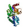
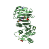
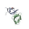
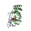
- PDB-4ygr: Crystal structure of HAD phosphatase from Thermococcus onnurineus -
+ Open data

- Basic information
| Entry | Database: PDB / ID: 4ygr | ||||||
|---|---|---|---|---|---|---|---|
| Title | Crystal structure of HAD phosphatase from Thermococcus onnurineus | ||||||
 Components Components | Hydrolase | ||||||
 Keywords Keywords | HYDROLASE / HAD phosphatase / substrate selectivity | ||||||
| Function / homology |  Function and homology informationN-acylneuraminate-9-phosphatase activity / N-acetylneuraminate biosynthetic process / dephosphorylation / metal ion binding Function and homology informationN-acylneuraminate-9-phosphatase activity / N-acetylneuraminate biosynthetic process / dephosphorylation / metal ion bindingSimilarity search - Function | ||||||
| Biological species |   Thermococcus onnurineus (archaea) Thermococcus onnurineus (archaea) | ||||||
| Method | X-RAY DIFFRACTION / SYNCHROTRON / Resolution: 1.703 Å | ||||||
 Authors Authors | Ngo, T.D. / Le, B.V. / Subramani, V.K. / Nguyen, C.M.T. / Lee, H.S. / Cho, Y. / Kim, K.K. / Hwang, H.Y. | ||||||
 Citation Citation | Journal: Biochem.Biophys.Res.Commun. / Year: 2015 Title: Structural basis for the substrate selectivity of a HAD phosphatase from Thermococcus onnurineus NA1 Authors: Ngo, T.D. / Le, B.V. / Subramani, V.K. / Nguyen, C.M.T. / Lee, H.S. / Cho, Y. / Kim, K.K. / Hwang, H.Y. | ||||||
| History |
|
- Structure visualization
| Structure viewer | Molecule: MolmilJmol/JSmol |
|---|
- Downloads & links
-Download
| PDBx/mmCIF format | 4ygr.cif.gz | 66.4 KB | Display | PDBx/mmCIF format |
|---|---|---|---|---|
| PDB format | pdb4ygr.ent.gz | 47.4 KB | Display | PDB format |
| PDBx/mmJSON format | 4ygr.json.gz | Tree view | PDBx/mmJSON format | |
| Others |  Other downloads Other downloads |
-Validation report
| Arichive directory | https://data.pdbj.org/pub/pdb/validation_reports/yg/4ygrftp://data.pdbj.org/pub/pdb/validation_reports/yg/4ygr | HTTPS FTP |
|---|
-Related structure data


-Links
 PDBj
PDBj

- Assembly
| Deposited unit | 
| ||||||||
|---|---|---|---|---|---|---|---|---|---|
| 1 |
| ||||||||
| Unit cell |
|
-Components
| #1: Protein | Mass: 26760.705 Da / Num. of mol.: 1 Source method: isolated from a genetically manipulated source Source: (gene. exp.) Thermococcus onnurineus (strain NA1) (archaea)Strain: NA1 / Gene: TON_0338 / Production host:  Escherichia coli (E. coli) / References: UniProt: B6YTD6 Escherichia coli (E. coli) / References: UniProt: B6YTD6 | ||||
|---|---|---|---|---|---|
| #2: Chemical | CHES (buffer)  Mass: 207.290 Da / Num. of mol.: 2 / Source method: obtained synthetically / Formula: C8H17NO3S / Comment: pH buffer*YM Mass: 207.290 Da / Num. of mol.: 2 / Source method: obtained synthetically / Formula: C8H17NO3S / Comment: pH buffer*YM#3: Chemical | ChemComp-MG / |   Mass: 24.305 Da / Num. of mol.: 1 / Source method: obtained synthetically / Formula: Mg Mass: 24.305 Da / Num. of mol.: 1 / Source method: obtained synthetically / Formula: Mg#4: Water | ChemComp-HOH / | Water Mass: 18.015 Da / Num. of mol.: 198 / Source method: isolated from a natural source / Formula: H2O Mass: 18.015 Da / Num. of mol.: 198 / Source method: isolated from a natural source / Formula: H2O |
-Experimental details
-Experiment
| Experiment | Method: X-RAY DIFFRACTION |
|---|
- Sample preparation
| Crystal | Density Matthews: 2.53 Å3/Da / Density % sol: 51.45 % |
|---|---|
| Crystal grow | Temperature: 287 K / Method: evaporation / pH: 9.5 / Details: 1.0M sodium citrate, 0.1M CHES (pH 9.5) |
-Data collection
| Diffraction | Mean temperature: 100 K |
|---|---|
| Diffraction source | Source: SYNCHROTRON / Site: PAL/PLS  / Beamline: 4A / Wavelength: 1 Å / Beamline: 4A / Wavelength: 1 Å |
| Detector | Type: ADSC QUANTUM 315 / Detector: CCD / Date: Mar 12, 2007 |
| Radiation | Protocol: SINGLE WAVELENGTH / Monochromatic (M) / Laue (L): M / Scattering type: x-ray |
| Radiation wavelength | Wavelength: 1 Å / Relative weight: 1 |
| Reflection | Resolution: 1.7→30 Å / Num. obs: 28848 / % possible obs: 98.3 % / Redundancy: 3.5 % / Net I/σ(I): 21.7 |
| Reflection shell | Resolution: 1.7→1.73 Å / Redundancy: 3.4 % / Rmerge(I) obs: 0.339 / Mean I/σ(I) obs: 2.7 / % possible all: 95.1 |
- Processing
| Software |
| |||||||||||||||||||||||||||||||||||||||||||||||||||||||||||||||||||||||||||||||||||||||||||||||||||||||||
|---|---|---|---|---|---|---|---|---|---|---|---|---|---|---|---|---|---|---|---|---|---|---|---|---|---|---|---|---|---|---|---|---|---|---|---|---|---|---|---|---|---|---|---|---|---|---|---|---|---|---|---|---|---|---|---|---|---|---|---|---|---|---|---|---|---|---|---|---|---|---|---|---|---|---|---|---|---|---|---|---|---|---|---|---|---|---|---|---|---|---|---|---|---|---|---|---|---|---|---|---|---|---|---|---|---|---|
| Refinement | Resolution: 1.703→29.242 Å / SU ML: 0.17 / Cross valid method: FREE R-VALUE / σ(F): 1.36 / Phase error: 18.42 / Stereochemistry target values: ML
| |||||||||||||||||||||||||||||||||||||||||||||||||||||||||||||||||||||||||||||||||||||||||||||||||||||||||
| Solvent computation | Shrinkage radii: 0.9 Å / VDW probe radii: 1.11 Å / Solvent model: FLAT BULK SOLVENT MODEL | |||||||||||||||||||||||||||||||||||||||||||||||||||||||||||||||||||||||||||||||||||||||||||||||||||||||||
| Refinement step | Cycle: LAST / Resolution: 1.703→29.242 Å
| |||||||||||||||||||||||||||||||||||||||||||||||||||||||||||||||||||||||||||||||||||||||||||||||||||||||||
| Refine LS restraints |
| |||||||||||||||||||||||||||||||||||||||||||||||||||||||||||||||||||||||||||||||||||||||||||||||||||||||||
| LS refinement shell |
|
