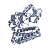
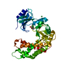
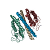
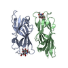
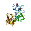
Entry Database : PDB / ID : 5axiTitle Crystal structure of Cbl-b TKB domain in complex with Cblin Cblin E3 ubiquitin-protein ligase CBL-B Keywords / / / / Function / homology Function Domain/homology Component
/ / / / / / / / / / / / / / / / / / / / / / / / / / / / / / / / / / / / / / / / / / / / / / / / / / / / / / / / / / / / / / / / / / / / / / / / / / / / / / / / / / / / / / / / / / / / / / / / / / / / / / / / / / / / / / / / / / / / / / / / / / / / / / / / / / / / / / / / / / / / / / / / Biological species Mus musculus (house mouse)Method / / / Resolution : 2.5 Å Authors Ohno, A. / Maita, N. / Ochi, A. / Nakao, R. / Nikawa, T. Journal : Arch.Biochem.Biophys. / Year : 2016Title : Structural analysis of the TKB domain of ubiquitin ligase Cbl-b complexed with its small inhibitory peptide, CblinAuthors : Ohno, A. / Ochi, A. / Maita, N. / Ueji, T. / Bando, A. / Nakao, R. / Hirasaka, K. / Abe, T. / Teshima-Kondo, S. / Nemoto, H. / Okumura, Y. / Higashibata, A. / Yano, S. / Tochio, H. / Nikawa, T. History Deposition Jul 29, 2015 Deposition site / Processing site Revision 1.0 Mar 2, 2016 Provider / Type Revision 1.1 Mar 9, 2016 Group Revision 1.2 Feb 26, 2020 Group / Database references / Derived calculationsCategory / diffrn_source / pdbx_struct_oper_listItem / _diffrn_source.pdbx_synchrotron_site / _pdbx_struct_oper_list.symmetry_operationRevision 1.3 Nov 8, 2023 Group Data collection / Database references ... Data collection / Database references / Derived calculations / Refinement description Category chem_comp_atom / chem_comp_bond ... chem_comp_atom / chem_comp_bond / database_2 / pdbx_initial_refinement_model / struct_conn / struct_conn_type Item _database_2.pdbx_DOI / _database_2.pdbx_database_accession ... _database_2.pdbx_DOI / _database_2.pdbx_database_accession / _struct_conn.conn_type_id / _struct_conn.id / _struct_conn.pdbx_dist_value / _struct_conn.pdbx_leaving_atom_flag / _struct_conn.ptnr1_auth_asym_id / _struct_conn.ptnr1_auth_comp_id / _struct_conn.ptnr1_auth_seq_id / _struct_conn.ptnr1_label_asym_id / _struct_conn.ptnr1_label_atom_id / _struct_conn.ptnr1_label_comp_id / _struct_conn.ptnr1_label_seq_id / _struct_conn.ptnr2_auth_asym_id / _struct_conn.ptnr2_auth_comp_id / _struct_conn.ptnr2_auth_seq_id / _struct_conn.ptnr2_label_asym_id / _struct_conn.ptnr2_label_atom_id / _struct_conn.ptnr2_label_comp_id / _struct_conn.ptnr2_label_seq_id / _struct_conn_type.id Revision 1.4 Nov 15, 2023 Group / Category / chem_comp_bond / Item / _chem_comp_bond.atom_id_2
Show all Show less
 Movie
Movie Controller


 Open data
 Basic information
 Components
 Keywords
 phosphopeptide / cblin / LIGASE-LIGASE inhibitor complex
 Function and homology information
 Authors
 Citation
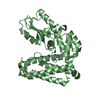
 Structure visualization
 Downloads & links
 Other downloads
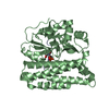


 PDBj
PDBj


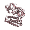
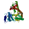
 Assembly


 Mass: 40.078 Da / Num. of mol.: 3 / Source method: obtained synthetically / Formula: Ca
 Mass: 35.453 Da / Num. of mol.: 4 / Source method: obtained synthetically / Formula: Cl
 Mass: 18.015 Da / Num. of mol.: 167 / Source method: isolated from a natural source / Formula: H2O
 Sample preparation
 / Beamline: AR-NW12A / Wavelength: 1 Å
 Processing