 Movie
Movie Controller

[English] 日本語
 Yorodumi
- PDB-4dbx: Crystal structure of aminoglycoside phosphotransferase APH(2")-ID... -
+ Open data

- Basic information
| Entry | Database: PDB / ID: 4dbx | |||||||||
|---|---|---|---|---|---|---|---|---|---|---|
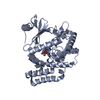
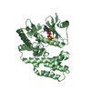
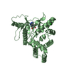
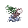
| Title | Crystal structure of aminoglycoside phosphotransferase APH(2")-ID/APH(2")-IVA | |||||||||
 Components Components | APH(2")-ID | |||||||||
 Keywords Keywords |  TRANSFERASE / STRUCTURAL GENOMICS / MCSG / Midwest Center for Structural Genomics / EUKARYOTIC PROTEIN KINASE-LIKE FOLD / AMINOGLYCOSIDE PHOSPHOTRANSFERASE / KINASE / AMINOGLYCOSIDES / INTRACELLULAR / PSI-Biology TRANSFERASE / STRUCTURAL GENOMICS / MCSG / Midwest Center for Structural Genomics / EUKARYOTIC PROTEIN KINASE-LIKE FOLD / AMINOGLYCOSIDE PHOSPHOTRANSFERASE / KINASE / AMINOGLYCOSIDES / INTRACELLULAR / PSI-Biology | |||||||||
| Function / homology |  Function and homology information Function and homology information | |||||||||
| Biological species |  Enterococcus casseliflavus (bacteria) Enterococcus casseliflavus (bacteria) | |||||||||
| Method | X-RAY DIFFRACTION / SYNCHROTRON / SAD / Resolution: 2.004 Å | |||||||||
 Authors Authors | Stogios, P.J. / Minasov, G. / Tan, K. / Nocek, B. / Singer, A.U. / Evdokimova, E. / Egorova, E. / Di Leo, R. / Li, H. / Shakya, T. ...Stogios, P.J. / Minasov, G. / Tan, K. / Nocek, B. / Singer, A.U. / Evdokimova, E. / Egorova, E. / Di Leo, R. / Li, H. / Shakya, T. / Wright, G.D. / Savchenko, A. / Anderson, W.F. / Midwest Center for Structural Genomics (MCSG) | |||||||||
 Citation Citation | Journal: Chem.Biol. / Year: 2011 Title: A small molecule discrimination map of the antibiotic resistance kinome. Authors: Shakya, T. / Stogios, P.J. / Waglechner, N. / Evdokimova, E. / Ejim, L. / Blanchard, J.E. / McArthur, A.G. / Savchenko, A. / Wright, G.D. | |||||||||
| History |
|
- Structure visualization
| Structure viewer | Molecule: MolmilJmol/JSmol |
|---|
- Downloads & links
-Download
| PDBx/mmCIF format | 4dbx.cif.gz | 135.1 KB | Display | PDBx/mmCIF format |
|---|---|---|---|---|
| PDB format | pdb4dbx.ent.gz | 110.5 KB | Display | PDB format |
| PDBx/mmJSON format | 4dbx.json.gz | Tree view | PDBx/mmJSON format | |
| Others |  Other downloads Other downloads |
-Validation report
| Arichive directory | https://data.pdbj.org/pub/pdb/validation_reports/db/4dbxftp://data.pdbj.org/pub/pdb/validation_reports/db/4dbx | HTTPS FTP |
|---|
-Related structure data
| Related structure data |  4de4C  4dfbC  4dfuC C: citing same article ( |
|---|---|
| Similar structure data | |
| Other databases |


-Links
 PDBj
PDBj
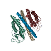
- Assembly
| Deposited unit | 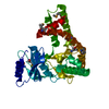
| ||||||||
|---|---|---|---|---|---|---|---|---|---|
| 1 |
| ||||||||
| Unit cell |
|
-Components
| #1: Protein | Mass: 38279.039 Da / Num. of mol.: 1 Source method: isolated from a genetically manipulated source Source: (gene. exp.) Enterococcus casseliflavus (bacteria) / Gene: APH(2")-ID / Plasmid: P15TV LIC / Production host: Escherichia coli (E. coli) / Strain (production host): BL21(DE3) / References: UniProt: O68183 |
|---|---|
| #2: Water | ChemComp-HOH / Water Mass: 18.015 Da / Num. of mol.: 300 / Source method: isolated from a natural source / Formula: H2O Mass: 18.015 Da / Num. of mol.: 300 / Source method: isolated from a natural source / Formula: H2O |
-Experimental details
-Experiment
| Experiment | Method: X-RAY DIFFRACTION / Number of used crystals: 1 |
|---|
- Sample preparation
| Crystal | Density Matthews: 2.42 Å3/Da / Density % sol: 49.08 % |
|---|---|
| Crystal grow | Temperature: 298 K / Method: vapor diffusion, sitting drop / pH: 7.5 Details: 0.1 M HEPES, 10% PEG 6K, 5% MPD, pH 7.5, VAPOR DIFFUSION, SITTING DROP, temperature 298K |
-Data collection
| Diffraction | Mean temperature: 100 K |
|---|---|
| Diffraction source | Source: SYNCHROTRON / Site: APS  / Beamline: 19-ID / Wavelength: 0.9794 Å / Beamline: 19-ID / Wavelength: 0.9794 Å |
| Detector | Type: ADSC QUANTUM 315r / Detector: CCD / Date: Aug 9, 2009 |
| Radiation | Monochromator: GRAPHITE / Protocol: SINGLE WAVELENGTH / Monochromatic (M) / Laue (L): M / Scattering type: x-ray |
| Radiation wavelength | Wavelength: 0.9794 Å / Relative weight: 1 |
| Reflection | Resolution: 2→50 Å / Num. obs: 25489 / % possible obs: 99.5 % / Observed criterion σ(F): 0 / Observed criterion σ(I): -3 / Redundancy: 7 % / Rsym value: 0.088 / Net I/σ(I): 34.04 |
| Reflection shell | Resolution: 2→2.07 Å / Redundancy: 5.9 % / Mean I/σ(I) obs: 5.13 / Rsym value: 0.508 / % possible all: 96.3 |
- Processing
| Software |
| |||||||||||||||||||||||||||||||||||||||||||||||||||||||||||||||||||||||||||
|---|---|---|---|---|---|---|---|---|---|---|---|---|---|---|---|---|---|---|---|---|---|---|---|---|---|---|---|---|---|---|---|---|---|---|---|---|---|---|---|---|---|---|---|---|---|---|---|---|---|---|---|---|---|---|---|---|---|---|---|---|---|---|---|---|---|---|---|---|---|---|---|---|---|---|---|---|
| Refinement | Method to determine structure: SAD / Resolution: 2.004→30.269 Å / SU ML: 0.18 / Cross valid method: THROUGHOUT / σ(F): 1.33 / Phase error: 21.16 / Stereochemistry target values: ML
| |||||||||||||||||||||||||||||||||||||||||||||||||||||||||||||||||||||||||||
| Solvent computation | Shrinkage radii: 0.9 Å / VDW probe radii: 1.11 Å / Solvent model: FLAT BULK SOLVENT MODEL / Bsol: 39.226 Å2 / ksol: 0.319 e/Å3 | |||||||||||||||||||||||||||||||||||||||||||||||||||||||||||||||||||||||||||
| Displacement parameters |
| |||||||||||||||||||||||||||||||||||||||||||||||||||||||||||||||||||||||||||
| Refinement step | Cycle: LAST / Resolution: 2.004→30.269 Å
| |||||||||||||||||||||||||||||||||||||||||||||||||||||||||||||||||||||||||||
| Refine LS restraints |
| |||||||||||||||||||||||||||||||||||||||||||||||||||||||||||||||||||||||||||
| LS refinement shell |
| |||||||||||||||||||||||||||||||||||||||||||||||||||||||||||||||||||||||||||
| Refinement TLS params. | Method: refined / Refine-ID: X-RAY DIFFRACTION
| |||||||||||||||||||||||||||||||||||||||||||||||||||||||||||||||||||||||||||
| Refinement TLS group |
|
