 Movie
Movie Controller

[English] 日本語
 Yorodumi
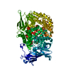
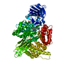
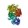
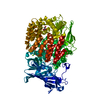
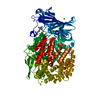
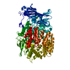
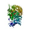
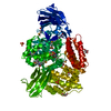
- PDB-4zx5: X-ray crystal structure of PfA-M1 in complex with hydroxamic acid... -
+ Open data

- Basic information
| Entry | Database: PDB / ID: 4zx5 | ||||||
|---|---|---|---|---|---|---|---|
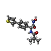
| Title | X-ray crystal structure of PfA-M1 in complex with hydroxamic acid-based inhibitor 10q | ||||||
 Components Components | M1 family aminopeptidase | ||||||
 Keywords Keywords | HYDROLASE/HYDROLASE INHIBITOR / M1 ALANYL-AMINOPEPTIDASE /  PROTEASE / INHIBITOR / HYDROXAMIC ACID / HYDROLASE-HYDROLASE INHIBITOR complex PROTEASE / INHIBITOR / HYDROXAMIC ACID / HYDROLASE-HYDROLASE INHIBITOR complex | ||||||
| Function / homology |  Function and homology information Function and homology informationsymbiont-containing vacuole membrane / vacuolar lumen / Hydrolases; Acting on peptide bonds (peptidases); Aminopeptidases / food vacuole / dipeptidase activity / metalloaminopeptidase activity / aminopeptidase activity / chloroplast / proteolysis / zinc ion binding ...symbiont-containing vacuole membrane / vacuolar lumen / Hydrolases; Acting on peptide bonds (peptidases); Aminopeptidases / food vacuole / dipeptidase activity / metalloaminopeptidase activity / aminopeptidase activity / chloroplast / proteolysis / zinc ion binding / membrane / nucleus / cytoplasmSimilarity search - Function | ||||||
| Biological species |  Plasmodium falciparum (malaria parasite P. falciparum) Plasmodium falciparum (malaria parasite P. falciparum) | ||||||
| Method | X-RAY DIFFRACTION / SYNCHROTRON / MOLECULAR REPLACEMENT / Resolution: 1.95 Å | ||||||
 Authors Authors | Drinkwater, N. / McGowan, S. | ||||||
| Funding support |  Australia, 1items Australia, 1items
| ||||||
 Citation Citation | Journal: Eur.J.Med.Chem. / Year: 2016 Title: Potent dual inhibitors of Plasmodium falciparum M1 and M17 aminopeptidases through optimization of S1 pocket interactions. Authors: Drinkwater, N. / Vinh, N.B. / Mistry, S.N. / Bamert, R.S. / Ruggeri, C. / Holleran, J.P. / Loganathan, S. / Paiardini, A. / Charman, S.A. / Powell, A.K. / Avery, V.M. / McGowan, S. / Scammells, P.J. | ||||||
| History |
|
- Structure visualization
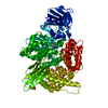
| Structure viewer | Molecule: MolmilJmol/JSmol |
|---|
- Downloads & links
-Download
| PDBx/mmCIF format | 4zx5.cif.gz | 419.8 KB | Display | PDBx/mmCIF format |
|---|---|---|---|---|
| PDB format | pdb4zx5.ent.gz | 340.2 KB | Display | PDB format |
| PDBx/mmJSON format | 4zx5.json.gz | Tree view | PDBx/mmJSON format | |
| Others |  Other downloads Other downloads |
-Validation report
| Arichive directory | https://data.pdbj.org/pub/pdb/validation_reports/zx/4zx5ftp://data.pdbj.org/pub/pdb/validation_reports/zx/4zx5 | HTTPS FTP |
|---|
-Related structure data
| Related structure data |  4zw3C  4zw5C  4zw6C 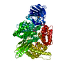 4zw7C 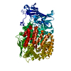 4zw8C  4zx3C  4zx4C  4zx6C 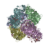 4zx8C 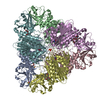 4zx9C  4zy0C 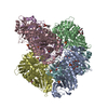 4zy1C 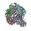 4zy2C 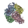 4zyqC  3ebgS S: Starting model for refinement C: citing same article ( |
|---|---|
| Similar structure data |


-Links
 PDBj
PDBj- Assembly
| Deposited unit | 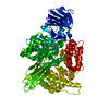
| ||||||||
|---|---|---|---|---|---|---|---|---|---|
| 1 |
| ||||||||
| Unit cell |
|
-Components
-Protein , 1 types, 1 molecules A
| #1: Protein | Mass: 103760.719 Da / Num. of mol.: 1 / Fragment: UNP residues 195-1084 / Mutation: N213Q, N223Q, H378P, N501Q, N795Q, N1069Q Source method: isolated from a genetically manipulated source Source: (gene. exp.) Plasmodium falciparum (malaria parasite P. falciparum)Strain: isolate FcB1 / Columbia / Plasmid: PTRCHIS-2B / Production host:  Escherichia coli (E. coli) / Strain (production host): BL21 Escherichia coli (E. coli) / Strain (production host): BL21References: UniProt: O96935, Hydrolases; Acting on peptide bonds (peptidases); Aminopeptidases |
|---|
-Non-polymers , 6 types, 987 molecules 


| #2: Chemical | ChemComp-ZN /  Mass: 65.409 Da / Num. of mol.: 1 / Source method: obtained synthetically / Formula: Zn Mass: 65.409 Da / Num. of mol.: 1 / Source method: obtained synthetically / Formula: Zn | ||||||
|---|---|---|---|---|---|---|---|
| #3: Chemical | ChemComp-4TM /  Mass: 332.417 Da / Num. of mol.: 1 / Source method: obtained synthetically / Formula: C17H20N2O3S Mass: 332.417 Da / Num. of mol.: 1 / Source method: obtained synthetically / Formula: C17H20N2O3S | ||||||
| #4: Chemical |  Mass: 24.305 Da / Num. of mol.: 2 / Source method: obtained synthetically / Formula: Mg Mass: 24.305 Da / Num. of mol.: 2 / Source method: obtained synthetically / Formula: Mg#5: Chemical | Dimethyl sulfoxide Mass: 78.133 Da / Num. of mol.: 2 / Source method: obtained synthetically / Formula: C2H6OS / Comment: DMSO, precipitant*YM Mass: 78.133 Da / Num. of mol.: 2 / Source method: obtained synthetically / Formula: C2H6OS / Comment: DMSO, precipitant*YM#6: Chemical | ChemComp-GOL / | Glycerol Mass: 92.094 Da / Num. of mol.: 1 / Source method: obtained synthetically / Formula: C3H8O3 Mass: 92.094 Da / Num. of mol.: 1 / Source method: obtained synthetically / Formula: C3H8O3#7: Water | ChemComp-HOH / | WaterMass: 18.015 Da / Num. of mol.: 980 / Source method: isolated from a natural source / Formula: H2O |
-Experimental details
-Experiment
| Experiment | Method: X-RAY DIFFRACTION / Number of used crystals: 1 |
|---|
- Sample preparation
| Crystal | Density Matthews: 2.31 Å3/Da / Density % sol: 46.87 % |
|---|---|
| Crystal grow | Temperature: 298 K / Method: vapor diffusion, hanging drop / pH: 8.5 Details: 22% (v/v) PEG 8000, 10% (v/v) glycerol, 0.1 M Tris pH 8.5, 0.2 M MgCl2 |
-Data collection
| Diffraction | Mean temperature: 100 K |
|---|---|
| Diffraction source | Source: SYNCHROTRON / Site: Australian Synchrotron / Beamline: MX1 / Wavelength: 0.9537 Å |
| Detector | Type: ADSC QUANTUM 210r / Detector: CCD / Date: Jul 26, 2014 |
| Radiation | Monochromator: DOUBLE CRYSTAL SILICON 111 / Protocol: SINGLE WAVELENGTH / Monochromatic (M) / Laue (L): M / Scattering type: x-ray |
| Radiation wavelength | Wavelength: 0.9537 Å / Relative weight: 1 |
| Reflection | Resolution: 1.95→31.56 Å / Num. obs: 70857 / % possible obs: 100 % / Redundancy: 7.3 % / Biso Wilson estimate: 21.31 Å2 / Rmerge(I) obs: 0.166 / Net I/σ(I): 11.1 |
| Reflection shell | Resolution: 1.95→1.99 Å / Redundancy: 7.3 % / Rmerge(I) obs: 1.276 / Mean I/σ(I) obs: 1.7 / % possible all: 100 |
- Processing
| Software |
| |||||||||||||||||||||||||||||||||||||||||||||||||||||||||||||||||||||||||||||||||||||||||||||||||||||||||||||||||||||||||||||||||||||||||||||||||||||||||||||||||||||||||||||||||||||||||||||
|---|---|---|---|---|---|---|---|---|---|---|---|---|---|---|---|---|---|---|---|---|---|---|---|---|---|---|---|---|---|---|---|---|---|---|---|---|---|---|---|---|---|---|---|---|---|---|---|---|---|---|---|---|---|---|---|---|---|---|---|---|---|---|---|---|---|---|---|---|---|---|---|---|---|---|---|---|---|---|---|---|---|---|---|---|---|---|---|---|---|---|---|---|---|---|---|---|---|---|---|---|---|---|---|---|---|---|---|---|---|---|---|---|---|---|---|---|---|---|---|---|---|---|---|---|---|---|---|---|---|---|---|---|---|---|---|---|---|---|---|---|---|---|---|---|---|---|---|---|---|---|---|---|---|---|---|---|---|---|---|---|---|---|---|---|---|---|---|---|---|---|---|---|---|---|---|---|---|---|---|---|---|---|---|---|---|---|---|---|---|---|
| Refinement | Method to determine structure: MOLECULAR REPLACEMENT Starting model: 3EBG Resolution: 1.95→31.56 Å / SU ML: 0.22 / Cross valid method: FREE R-VALUE / σ(F): 1.34 / Phase error: 19.82 / Stereochemistry target values: ML
| |||||||||||||||||||||||||||||||||||||||||||||||||||||||||||||||||||||||||||||||||||||||||||||||||||||||||||||||||||||||||||||||||||||||||||||||||||||||||||||||||||||||||||||||||||||||||||||
| Solvent computation | Shrinkage radii: 0.9 Å / VDW probe radii: 1.11 Å / Solvent model: FLAT BULK SOLVENT MODEL | |||||||||||||||||||||||||||||||||||||||||||||||||||||||||||||||||||||||||||||||||||||||||||||||||||||||||||||||||||||||||||||||||||||||||||||||||||||||||||||||||||||||||||||||||||||||||||||
| Displacement parameters | Biso mean: 25.46 Å2 | |||||||||||||||||||||||||||||||||||||||||||||||||||||||||||||||||||||||||||||||||||||||||||||||||||||||||||||||||||||||||||||||||||||||||||||||||||||||||||||||||||||||||||||||||||||||||||||
| Refinement step | Cycle: LAST / Resolution: 1.95→31.56 Å
| |||||||||||||||||||||||||||||||||||||||||||||||||||||||||||||||||||||||||||||||||||||||||||||||||||||||||||||||||||||||||||||||||||||||||||||||||||||||||||||||||||||||||||||||||||||||||||||
| Refine LS restraints |
| |||||||||||||||||||||||||||||||||||||||||||||||||||||||||||||||||||||||||||||||||||||||||||||||||||||||||||||||||||||||||||||||||||||||||||||||||||||||||||||||||||||||||||||||||||||||||||||
| LS refinement shell |
| |||||||||||||||||||||||||||||||||||||||||||||||||||||||||||||||||||||||||||||||||||||||||||||||||||||||||||||||||||||||||||||||||||||||||||||||||||||||||||||||||||||||||||||||||||||||||||||
| Refinement TLS params. | Method: refined / Origin x: 92.3071 Å / Origin y: 113.0077 Å / Origin z: 10.2482 Å
| |||||||||||||||||||||||||||||||||||||||||||||||||||||||||||||||||||||||||||||||||||||||||||||||||||||||||||||||||||||||||||||||||||||||||||||||||||||||||||||||||||||||||||||||||||||||||||||
| Refinement TLS group | Selection details: ALL |
