 Movie
Movie Controller

[English] 日本語
 Yorodumi
- PDB-4r5t: Structure of the m1 alanylaminopeptidase from malaria complexed w... -
+ Open data

- Basic information
| Entry | Database: PDB / ID: 4r5t | ||||||
|---|---|---|---|---|---|---|---|
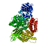
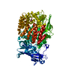
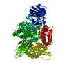
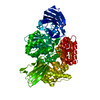
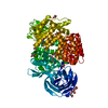
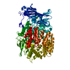
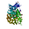
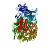
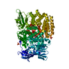
| Title | Structure of the m1 alanylaminopeptidase from malaria complexed with a hydroxamic acid-based inhibitor | ||||||
 Components Components | M1 family aminopeptidase | ||||||
 Keywords Keywords | HYDROLASE/HYDROLASE INHIBITOR /  PROTEASE / HYDROLASE-HYDROLASE INHIBITOR complex PROTEASE / HYDROLASE-HYDROLASE INHIBITOR complex | ||||||
| Function / homology |  Function and homology information Function and homology informationsymbiont-containing vacuole membrane / vacuolar lumen / Hydrolases; Acting on peptide bonds (peptidases); Aminopeptidases / food vacuole / dipeptidase activity / metalloaminopeptidase activity / aminopeptidase activity / chloroplast / proteolysis / zinc ion binding ...symbiont-containing vacuole membrane / vacuolar lumen / Hydrolases; Acting on peptide bonds (peptidases); Aminopeptidases / food vacuole / dipeptidase activity / metalloaminopeptidase activity / aminopeptidase activity / chloroplast / proteolysis / zinc ion binding / membrane / nucleus / cytoplasmSimilarity search - Function | ||||||
| Biological species |  Plasmodium falciparum FcB1/Columbia (eukaryote) Plasmodium falciparum FcB1/Columbia (eukaryote) | ||||||
| Method | X-RAY DIFFRACTION / SYNCHROTRON / MOLECULAR REPLACEMENT / molecular replacement / Resolution: 1.98 Å | ||||||
 Authors Authors | Drinkwater, N. / Mcgowan, S. | ||||||
 Citation Citation | Journal: J.Med.Chem. / Year: 2014 Title: Two-Pronged Attack: Dual Inhibition of Plasmodium falciparum M1 and M17 Metalloaminopeptidases by a Novel Series of Hydroxamic Acid-Based Inhibitors. Authors: Mistry, S.N. / Drinkwater, N. / Ruggeri, C. / Sivaraman, K.K. / Loganathan, S. / Fletcher, S. / Drag, M. / Paiardini, A. / Avery, V.M. / Scammells, P.J. / McGowan, S. | ||||||
| History |
|
- Structure visualization
| Structure viewer | Molecule: MolmilJmol/JSmol |
|---|
- Downloads & links
-Download
| PDBx/mmCIF format | 4r5t.cif.gz | 395.5 KB | Display | PDBx/mmCIF format |
|---|---|---|---|---|
| PDB format | pdb4r5t.ent.gz | 316.9 KB | Display | PDB format |
| PDBx/mmJSON format | 4r5t.json.gz | Tree view | PDBx/mmJSON format | |
| Others |  Other downloads Other downloads |
-Validation report
| Arichive directory | https://data.pdbj.org/pub/pdb/validation_reports/r5/4r5tftp://data.pdbj.org/pub/pdb/validation_reports/r5/4r5t | HTTPS FTP |
|---|
-Related structure data
| Related structure data |  4r5vC 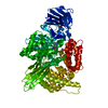 4r5xC 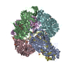 4r6tC  4r76C 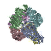 4r7mC  3ebgS S: Starting model for refinement C: citing same article ( |
|---|---|
| Similar structure data |


-Links
 PDBj
PDBj- Assembly
| Deposited unit | 
| ||||||||
|---|---|---|---|---|---|---|---|---|---|
| 1 |
| ||||||||
| Unit cell |
| ||||||||
| Details | biological unit is the same as asym. |
-Components
-Protein , 1 types, 1 molecules A
| #1: Protein | Mass: 104598.641 Da / Num. of mol.: 1 / Fragment: UNP residues 195-1084 Source method: isolated from a genetically manipulated source Source: (gene. exp.) Plasmodium falciparum FcB1/Columbia (eukaryote)Plasmid: PTRCHIS-2B / Production host:  Escherichia coli (E. coli) / Strain (production host): BL21 Escherichia coli (E. coli) / Strain (production host): BL21References: UniProt: O96935, Hydrolases; Acting on peptide bonds (peptidases); Aminopeptidases |
|---|
-Non-polymers , 5 types, 491 molecules 


| #2: Chemical | ChemComp-ZN /  Mass: 65.409 Da / Num. of mol.: 1 / Source method: obtained synthetically / Formula: Zn Mass: 65.409 Da / Num. of mol.: 1 / Source method: obtained synthetically / Formula: Zn | ||||||
|---|---|---|---|---|---|---|---|
| #3: Chemical | Dimethyl sulfoxide Mass: 78.133 Da / Num. of mol.: 2 / Source method: obtained synthetically / Formula: C2H6OS / Comment: DMSO, precipitant*YM Mass: 78.133 Da / Num. of mol.: 2 / Source method: obtained synthetically / Formula: C2H6OS / Comment: DMSO, precipitant*YM#4: Chemical | ChemComp-GOL / Glycerol Mass: 92.094 Da / Num. of mol.: 8 / Source method: obtained synthetically / Formula: C3H8O3 Mass: 92.094 Da / Num. of mol.: 8 / Source method: obtained synthetically / Formula: C3H8O3#5: Chemical | ChemComp-R5T / |  Mass: 332.354 Da / Num. of mol.: 1 / Source method: obtained synthetically / Formula: C16H20N4O4 Mass: 332.354 Da / Num. of mol.: 1 / Source method: obtained synthetically / Formula: C16H20N4O4#6: Water | ChemComp-HOH / | WaterMass: 18.015 Da / Num. of mol.: 479 / Source method: isolated from a natural source / Formula: H2O |
-Experimental details
-Experiment
| Experiment | Method: X-RAY DIFFRACTION / Number of used crystals: 1 |
|---|
- Sample preparation
| Crystal | Density Matthews: 2.33 Å3/Da / Density % sol: 47.29 % |
|---|---|
| Crystal grow | Temperature: 298 K / Method: vapor diffusion, hanging drop / pH: 8.5 Details: 22% (v/v) PEG 8000, 10* (v/v) glycerol, 0.1 M Tris pH 8.5, 0.2 M MgCl2, vapor diffusion, hanging drop, temperature 298K |
-Data collection
| Diffraction | Mean temperature: 100 K | ||||||||||||||||||
|---|---|---|---|---|---|---|---|---|---|---|---|---|---|---|---|---|---|---|---|
| Diffraction source | Source: SYNCHROTRON / Site: Australian Synchrotron  / Beamline: MX1 / Wavelength: 0.95369 Å / Beamline: MX1 / Wavelength: 0.95369 Å | ||||||||||||||||||
| Detector | Type: ADSC QUANTUM 210r / Detector: CCD / Date: Jun 12, 2013 | ||||||||||||||||||
| Radiation | Monochromator: DOUBLE CRYSTAL SILICON 111 / Protocol: SINGLE WAVELENGTH / Monochromatic (M) / Laue (L): M / Scattering type: x-ray | ||||||||||||||||||
| Radiation wavelength | Wavelength: 0.95369 Å / Relative weight: 1 | ||||||||||||||||||
| Reflection | Resolution: 1.98→37.6 Å / Num. all: 68739 / Num. obs: 68739 / % possible obs: 99.7 % / Redundancy: 20.6 % / Rmerge(I) obs: 0.169 / Net I/σ(I): 12.7 / Scaling rejects: 176 | ||||||||||||||||||
| Reflection shell | Rmerge(I) obs: 0.02 / Diffraction-ID: 1
|
-Phasing
| Phasing | Method: molecular replacement |
|---|
- Processing
| Software |
| ||||||||||||||||||||||||||||||||||||||||
|---|---|---|---|---|---|---|---|---|---|---|---|---|---|---|---|---|---|---|---|---|---|---|---|---|---|---|---|---|---|---|---|---|---|---|---|---|---|---|---|---|---|
| Refinement | Method to determine structure: MOLECULAR REPLACEMENT Starting model: 3EBG Resolution: 1.98→35.8418 Å / σ(F): 1.5 / Stereochemistry target values: Engh & Huber
| ||||||||||||||||||||||||||||||||||||||||
| Displacement parameters | Biso max: 105.18 Å2 / Biso mean: 40.491 Å2 / Biso min: 12.57 Å2 | ||||||||||||||||||||||||||||||||||||||||
| Refinement step | Cycle: LAST / Resolution: 1.98→35.8418 Å
| ||||||||||||||||||||||||||||||||||||||||
| Refinement TLS params. | Method: refined / Origin x: 17.5233 Å / Origin y: 3.6062 Å / Origin z: 10.1864 Å
| ||||||||||||||||||||||||||||||||||||||||
| Refinement TLS group |
|
