 Movie
Movie Controller

[English] 日本語
 Yorodumi
- PDB-4yry: Insights into flavin-based electron bifurcation via the NADH-depe... -
+ Open data

- Basic information
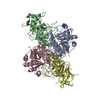
| Entry | Database: PDB / ID: 4yry | ||||||
|---|---|---|---|---|---|---|---|
| Title | Insights into flavin-based electron bifurcation via the NADH-dependent reduced ferredoxin-NADP oxidoreductase structure | ||||||
 Components Components |
| ||||||
 Keywords Keywords |  OXIDOREDUCTASE OXIDOREDUCTASE | ||||||
| Function / homology |  Function and homology information Function and homology informationpyrimidine nucleotide biosynthetic process / 2 iron, 2 sulfur cluster binding / flavin adenine dinucleotide binding / 4 iron, 4 sulfur cluster binding / oxidoreductase activity / nucleotide binding / metal ion bindingSimilarity search - Function | ||||||
| Biological species |   Thermotoga maritima (bacteria) Thermotoga maritima (bacteria) | ||||||
| Method | X-RAY DIFFRACTION / SYNCHROTRON / MOLECULAR REPLACEMENT / Resolution: 2.4 Å | ||||||
 Authors Authors | Ermler, U. / Thauer, R.K. / Demmer, J.K. / Huang, H. / Wang, S. / Demmer, U. | ||||||
 Citation Citation | Journal: J.Biol.Chem. / Year: 2015 Title: Insights into Flavin-based Electron Bifurcation via the NADH-dependent Reduced Ferredoxin:NADP Oxidoreductase Structure. Authors: Demmer, J.K. / Huang, H. / Wang, S. / Demmer, U. / Thauer, R.K. / Ermler, U. | ||||||
| History |
|
- Structure visualization
| Structure viewer | Molecule: MolmilJmol/JSmol |
|---|
- Downloads & links
-Download
| PDBx/mmCIF format | 4yry.cif.gz | 589 KB | Display | PDBx/mmCIF format |
|---|---|---|---|---|
| PDB format | pdb4yry.ent.gz | 489.7 KB | Display | PDB format |
| PDBx/mmJSON format | 4yry.json.gz | Tree view | PDBx/mmJSON format | |
| Others |  Other downloads Other downloads |
-Validation report
| Arichive directory | https://data.pdbj.org/pub/pdb/validation_reports/yr/4yryftp://data.pdbj.org/pub/pdb/validation_reports/yr/4yry | HTTPS FTP |
|---|
-Related structure data
| Related structure data | 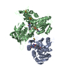 4ylfSC S: Starting model for refinement C: citing same article ( |
|---|---|
| Similar structure data |


-Links
 PDBj
PDBj


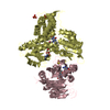
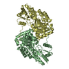
- Assembly
| Deposited unit | 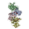
| ||||||||
|---|---|---|---|---|---|---|---|---|---|
| 1 | 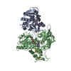
| ||||||||
| 2 | 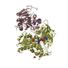
| ||||||||
| Unit cell |
|
-Components
-Protein , 2 types, 4 molecules ACBD
| #1: Protein | Mass: 30555.508 Da / Num. of mol.: 2 Source method: isolated from a genetically manipulated source Source: (gene. exp.) Thermotoga maritima (bacteria) / Gene: TM_1639 / Plasmid: pET-51b(+) / Production host: Escherichia coli BL21(DE3) (bacteria) / Variant (production host): C41 / References: UniProt: Q9X1X4#2: Protein | Mass: 51682.293 Da / Num. of mol.: 2 Source method: isolated from a genetically manipulated source Source: (gene. exp.) Thermotoga maritima (bacteria) / Gene: THEMA_06045 / Production host: Escherichia coli (E. coli) / Strain (production host): BL21(DE3) / Variant (production host): C41 / References: UniProt: V9X7T9, UniProt: Q9X1X5*PLUS |
|---|
-Non-polymers , 6 types, 145 molecules 


| #3: Chemical | Iron–sulfur cluster Mass: 175.820 Da / Num. of mol.: 2 / Source method: obtained synthetically / Formula: Fe2S2 Mass: 175.820 Da / Num. of mol.: 2 / Source method: obtained synthetically / Formula: Fe2S2#4: Chemical | ChemComp-FAD / Flavin adenine dinucleotide Mass: 785.550 Da / Num. of mol.: 4 / Source method: obtained synthetically / Formula: C27H33N9O15P2 / Comment: FAD*YM Mass: 785.550 Da / Num. of mol.: 4 / Source method: obtained synthetically / Formula: C27H33N9O15P2 / Comment: FAD*YM#5: Chemical | Nicotinamide adenine dinucleotide Mass: 663.425 Da / Num. of mol.: 2 / Source method: obtained synthetically / Formula: C21H27N7O14P2 / Comment: NAD*YM Mass: 663.425 Da / Num. of mol.: 2 / Source method: obtained synthetically / Formula: C21H27N7O14P2 / Comment: NAD*YM#6: Chemical | ChemComp-SF4 / Iron–sulfur cluster Mass: 351.640 Da / Num. of mol.: 4 / Source method: obtained synthetically / Formula: Fe4S4 Mass: 351.640 Da / Num. of mol.: 4 / Source method: obtained synthetically / Formula: Fe4S4#7: Chemical | ChemComp-PO4 / | Phosphate Mass: 94.971 Da / Num. of mol.: 1 / Source method: isolated from a natural source / Formula: PO4 Mass: 94.971 Da / Num. of mol.: 1 / Source method: isolated from a natural source / Formula: PO4#8: Water | ChemComp-HOH / | WaterMass: 18.015 Da / Num. of mol.: 132 / Source method: isolated from a natural source / Formula: H2O |
|---|
-Experimental details
-Experiment
| Experiment | Method: X-RAY DIFFRACTION |
|---|
- Sample preparation
| Crystal | Density Matthews: 3.1 Å3/Da / Density % sol: 60.27 % |
|---|---|
| Crystal grow | Temperature: 293.15 K / Method: vapor diffusion, hanging drop Details: 0.225M NH4H2PO4, 5 % (v,v) Ethanol, 20 % glycerol. soaked for 40 min in 5 mM NADH |
-Data collection
| Diffraction | Mean temperature: 100 K |
|---|---|
| Diffraction source | Source: SYNCHROTRON / Site: SLS  / Beamline: X10SA / Wavelength: 1 Å / Beamline: X10SA / Wavelength: 1 Å |
| Detector | Type: PSI PILATUS 6M / Detector: PIXEL / Date: Apr 9, 2013 |
| Radiation | Protocol: SINGLE WAVELENGTH / Monochromatic (M) / Laue (L): M / Scattering type: x-ray |
| Radiation wavelength | Wavelength: 1 Å / Relative weight: 1 |
| Reflection | Resolution: 2.4→50 Å / Num. obs: 76947 / % possible obs: 99.1 % / Redundancy: 5.2 % / Rsym value: 0.113 / Net I/σ(I): 14.2 |
| Reflection shell | Resolution: 2.4→2.5 Å / Redundancy: 5 % / Mean I/σ(I) obs: 2.3 / Rsym value: 0.896 / % possible all: 97.1 |
- Processing
| Software |
| |||||||||||||||||||||||||||||||||||||||||||||||||||||||||||||||||||||||||||||||||||||||||||||||||||||||||||||||||||||||||||||||||||||||||||||||||||
|---|---|---|---|---|---|---|---|---|---|---|---|---|---|---|---|---|---|---|---|---|---|---|---|---|---|---|---|---|---|---|---|---|---|---|---|---|---|---|---|---|---|---|---|---|---|---|---|---|---|---|---|---|---|---|---|---|---|---|---|---|---|---|---|---|---|---|---|---|---|---|---|---|---|---|---|---|---|---|---|---|---|---|---|---|---|---|---|---|---|---|---|---|---|---|---|---|---|---|---|---|---|---|---|---|---|---|---|---|---|---|---|---|---|---|---|---|---|---|---|---|---|---|---|---|---|---|---|---|---|---|---|---|---|---|---|---|---|---|---|---|---|---|---|---|---|---|---|---|
| Refinement | Method to determine structure: MOLECULAR REPLACEMENT Starting model: 4YLF Resolution: 2.4→19.96 Å / Cross valid method: FREE R-VALUE / σ(F): 1.37 / Phase error: 27.44 / Stereochemistry target values: TWIN_LSQ_F
| |||||||||||||||||||||||||||||||||||||||||||||||||||||||||||||||||||||||||||||||||||||||||||||||||||||||||||||||||||||||||||||||||||||||||||||||||||
| Solvent computation | Shrinkage radii: 0.9 Å / VDW probe radii: 1.11 Å / Solvent model: FLAT BULK SOLVENT MODEL | |||||||||||||||||||||||||||||||||||||||||||||||||||||||||||||||||||||||||||||||||||||||||||||||||||||||||||||||||||||||||||||||||||||||||||||||||||
| Refinement step | Cycle: LAST / Resolution: 2.4→19.96 Å
| |||||||||||||||||||||||||||||||||||||||||||||||||||||||||||||||||||||||||||||||||||||||||||||||||||||||||||||||||||||||||||||||||||||||||||||||||||
| Refine LS restraints |
| |||||||||||||||||||||||||||||||||||||||||||||||||||||||||||||||||||||||||||||||||||||||||||||||||||||||||||||||||||||||||||||||||||||||||||||||||||
| LS refinement shell |
| |||||||||||||||||||||||||||||||||||||||||||||||||||||||||||||||||||||||||||||||||||||||||||||||||||||||||||||||||||||||||||||||||||||||||||||||||||
| Refinement TLS params. | Method: refined / Refine-ID: X-RAY DIFFRACTION
| |||||||||||||||||||||||||||||||||||||||||||||||||||||||||||||||||||||||||||||||||||||||||||||||||||||||||||||||||||||||||||||||||||||||||||||||||||
| Refinement TLS group |
|
