 Movie
Movie Controller


+ Open data

- Basic information
| Entry | Database: PDB / ID: 4yhv | |||||||||
|---|---|---|---|---|---|---|---|---|---|---|
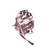
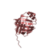
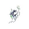
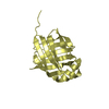
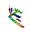
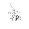
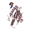
| Title | Yeast Prp3 C-terminal fragment 325-469 | |||||||||
 Components Components | U4/U6 small nuclear ribonucleoprotein PRP3 | |||||||||
 Keywords Keywords |  RNA BINDING PROTEIN / Spliceosomal protein / DUF1115 / RNA-binding domain / ferredoxin-like fold RNA BINDING PROTEIN / Spliceosomal protein / DUF1115 / RNA-binding domain / ferredoxin-like fold | |||||||||
| Function / homology |  Function and homology information Function and homology informationU4/U6 snRNP / U4 snRNP / spliceosomal complex assembly / spliceosomal snRNP assembly / U4/U6 x U5 tri-snRNP complex / spliceosomal complex / mRNA splicing, via spliceosome / nucleusSimilarity search - Function | |||||||||
| Biological species |  Saccharomyces cerevisiae (brewer's yeast) Saccharomyces cerevisiae (brewer's yeast) | |||||||||
| Method | X-RAY DIFFRACTION / SYNCHROTRON / MOLECULAR REPLACEMENT / Resolution: 2 Å | |||||||||
 Authors Authors | Liu, S. / Wahl, M.C. | |||||||||
| Funding support |  Germany, 1items Germany, 1items
| |||||||||
 Citation Citation | Journal: Elife / Year: 2015 Title: A composite double-/single-stranded RNA-binding region in protein Prp3 supports tri-snRNP stability and splicing. Authors: Liu, S. / Mozaffari-Jovin, S. / Wollenhaupt, J. / Santos, K.F. / Theuser, M. / Dunin-Horkawicz, S. / Fabrizio, P. / Bujnicki, J.M. / Luhrmann, R. / Wahl, M.C. | |||||||||
| History |
|
- Structure visualization
| Structure viewer | Molecule: MolmilJmol/JSmol |
|---|
- Downloads & links
-Download
| PDBx/mmCIF format | 4yhv.cif.gz | 75.1 KB | Display | PDBx/mmCIF format |
|---|---|---|---|---|
| PDB format | pdb4yhv.ent.gz | 55.2 KB | Display | PDB format |
| PDBx/mmJSON format | 4yhv.json.gz | Tree view | PDBx/mmJSON format | |
| Others |  Other downloads Other downloads |
-Validation report
| Arichive directory | https://data.pdbj.org/pub/pdb/validation_reports/yh/4yhvftp://data.pdbj.org/pub/pdb/validation_reports/yh/4yhv | HTTPS FTP |
|---|
-Related structure data
| Related structure data |  4yhuSC 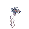 4yhwC S: Starting model for refinement C: citing same article ( |
|---|---|
| Similar structure data |


-Links
 PDBj
PDBj

- Assembly
| Deposited unit | 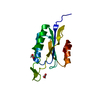
| ||||||||
|---|---|---|---|---|---|---|---|---|---|
| 1 |
| ||||||||
| Unit cell |
|
-Components
| #1: Protein | Mass: 17361.094 Da / Num. of mol.: 1 / Fragment: C-terminal fragment, UNP residues 325-469 Source method: isolated from a genetically manipulated source Source: (gene. exp.) Saccharomyces cerevisiae (brewer's yeast)Gene: PRP3, RNA3, YDR473C, D8035.16 / Production host:  Escherichia coli (E. coli) / References: UniProt: Q03338 Escherichia coli (E. coli) / References: UniProt: Q03338 | ||
|---|---|---|---|
| #2: Chemical | Acetic acid  Mass: 60.052 Da / Num. of mol.: 2 / Source method: obtained synthetically / Formula: C2H4O2 Mass: 60.052 Da / Num. of mol.: 2 / Source method: obtained synthetically / Formula: C2H4O2#3: Water | ChemComp-HOH / | Water Mass: 18.015 Da / Num. of mol.: 67 / Source method: isolated from a natural source / Formula: H2O Mass: 18.015 Da / Num. of mol.: 67 / Source method: isolated from a natural source / Formula: H2O |
-Experimental details
-Experiment
| Experiment | Method: X-RAY DIFFRACTION |
|---|
- Sample preparation
| Crystal | Density Matthews: 2.27 Å3/Da / Density % sol: 45.77 % |
|---|---|
| Crystal grow | Temperature: 291 K / Method: vapor diffusion, sitting drop / Details: 0.1 M HEPES, pH 7.5, 10 % PEG 8000 |
-Data collection
| Diffraction | Mean temperature: 100 K |
|---|---|
| Diffraction source | Source: SYNCHROTRON / Site: BESSY / Beamline: 14.2 / Wavelength: 0.91841 Å |
| Detector | Type: RAYONIX MX-225 / Detector: CCD / Date: May 30, 2012 |
| Radiation | Protocol: SINGLE WAVELENGTH / Monochromatic (M) / Laue (L): M / Scattering type: x-ray |
| Radiation wavelength | Wavelength: 0.91841 Å / Relative weight: 1 |
| Reflection | Resolution: 2→50 Å / Num. obs: 10459 / % possible obs: 99.3 % / Redundancy: 11.4 % / Rmerge(I) obs: 0.068 / Net I/σ(I): 24.96 |
| Reflection shell | Resolution: 2→2.12 Å / Redundancy: 11.3 % / Rmerge(I) obs: 0.439 / Mean I/σ(I) obs: 5.94 / % possible all: 96.2 |
- Processing
| Software |
| ||||||||||||||||||||||||||||||||||||||||||||||||||||||||||||||||||||||||||||||||||||||||||||||||||||
|---|---|---|---|---|---|---|---|---|---|---|---|---|---|---|---|---|---|---|---|---|---|---|---|---|---|---|---|---|---|---|---|---|---|---|---|---|---|---|---|---|---|---|---|---|---|---|---|---|---|---|---|---|---|---|---|---|---|---|---|---|---|---|---|---|---|---|---|---|---|---|---|---|---|---|---|---|---|---|---|---|---|---|---|---|---|---|---|---|---|---|---|---|---|---|---|---|---|---|---|---|---|
| Refinement | Method to determine structure: MOLECULAR REPLACEMENT Starting model: 4YHU Resolution: 2→24.283 Å / SU ML: 0.21 / Cross valid method: FREE R-VALUE / σ(F): 2 / Phase error: 22.34 / Stereochemistry target values: ML
| ||||||||||||||||||||||||||||||||||||||||||||||||||||||||||||||||||||||||||||||||||||||||||||||||||||
| Solvent computation | Shrinkage radii: 0.9 Å / VDW probe radii: 1.11 Å / Solvent model: FLAT BULK SOLVENT MODEL | ||||||||||||||||||||||||||||||||||||||||||||||||||||||||||||||||||||||||||||||||||||||||||||||||||||
| Refinement step | Cycle: LAST / Resolution: 2→24.283 Å
| ||||||||||||||||||||||||||||||||||||||||||||||||||||||||||||||||||||||||||||||||||||||||||||||||||||
| Refine LS restraints |
| ||||||||||||||||||||||||||||||||||||||||||||||||||||||||||||||||||||||||||||||||||||||||||||||||||||
| LS refinement shell |
| ||||||||||||||||||||||||||||||||||||||||||||||||||||||||||||||||||||||||||||||||||||||||||||||||||||
| Refinement TLS params. | Method: refined / Refine-ID: X-RAY DIFFRACTION
| ||||||||||||||||||||||||||||||||||||||||||||||||||||||||||||||||||||||||||||||||||||||||||||||||||||
| Refinement TLS group |
|
