 Movie
Movie Controller

[English] 日本語
 Yorodumi
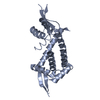
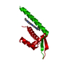
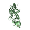
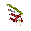
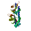
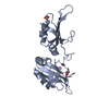
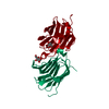
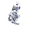
- PDB-4zzd: CRYSTAL STRUCTURE OF MULTIDRUG RESISTANCE REGULATOR LMRR BOUND TO... -
+ Open data

- Basic information
| Entry | Database: PDB / ID: 4zzd | ||||||
|---|---|---|---|---|---|---|---|
| Title | CRYSTAL STRUCTURE OF MULTIDRUG RESISTANCE REGULATOR LMRR BOUND TO RIBOFLAVIN | ||||||
 Components Components | TRANSCRIPTIONAL REGULATOR, PADR-LIKE FAMILY Transcriptional regulation Transcriptional regulation | ||||||
 Keywords Keywords | TRANSCRIPTION / WINGED HELIX TURN HELIX / TRANSCRIPTION REGULATOR / MULTIDRUG BINDING / INTRACELLULAR | ||||||
| Function / homology |  Function and homology information Function and homology informationTranscription regulator PadR, N-terminal / Transcriptional regulator PadR-like family / Winged helix-like DNA-binding domain superfamily/Winged helix DNA-binding domain / Arc Repressor Mutant, subunit A / Winged helix DNA-binding domain superfamily / Winged helix-like DNA-binding domain superfamily / Orthogonal Bundle / Mainly Alpha Similarity search - Domain/homology | ||||||
| Biological species |  Lactococcus lactis subsp. cremoris MG1363 (lactic acid bacteria) Lactococcus lactis subsp. cremoris MG1363 (lactic acid bacteria) | ||||||
| Method | X-RAY DIFFRACTION / SYNCHROTRON / MOLECULAR REPLACEMENT / Resolution: 2.351 Å | ||||||
 Authors Authors | Madoori, P.K. / Thunnissen, A.-M.W.H. | ||||||
 Citation Citation | Journal: Plos One / Year: 2015 Title: Binding of the Lactococcal Drug Dependent Transcriptional Regulator LmrR to Its Ligands and Responsive Promoter Regions. Authors: van der Berg, J.P. / Madoori, P.K. / Komarudin, A.G. / Thunnissen, A.M. / Driessen, A.J. | ||||||
| History |
|
- Structure visualization
| Structure viewer | Molecule: MolmilJmol/JSmol |
|---|
- Downloads & links
-Download
| PDBx/mmCIF format | 4zzd.cif.gz | 57.9 KB | Display | PDBx/mmCIF format |
|---|---|---|---|---|
| PDB format | pdb4zzd.ent.gz | 41.4 KB | Display | PDB format |
| PDBx/mmJSON format | 4zzd.json.gz | Tree view | PDBx/mmJSON format | |
| Others |  Other downloads Other downloads |
-Validation report
| Arichive directory | https://data.pdbj.org/pub/pdb/validation_reports/zz/4zzdftp://data.pdbj.org/pub/pdb/validation_reports/zz/4zzd | HTTPS FTP |
|---|
-Related structure data
| Related structure data | |
|---|---|
| Similar structure data |


-Links
 PDBj
PDBj- Assembly
| Deposited unit | 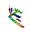
| |||||||||
|---|---|---|---|---|---|---|---|---|---|---|
| 1 | 
| |||||||||
| Unit cell |
| |||||||||
| Symmetry | Point symmetry: (Schoenflies symbol: C2 (2 fold cyclic)) | |||||||||
| Components on special symmetry positions |
|
-Components
| #1: Protein | Transcriptional regulation Mass: 13478.371 Da / Num. of mol.: 1 Source method: isolated from a genetically manipulated source Source: (gene. exp.) Lactococcus lactis subsp. cremoris MG1363 (lactic acid bacteria)Gene: llmg_0323 / Plasmid: PNSC8048 Production host: Lactococcus lactis subsp. cremoris NZ9000 (lactic acid bacteria)References: UniProt: A2RI36 |
|---|---|
| #2: Chemical | ChemComp-RBF / Riboflavin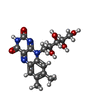  Mass: 376.364 Da / Num. of mol.: 1 / Source method: obtained synthetically / Formula: C17H20N4O6 Mass: 376.364 Da / Num. of mol.: 1 / Source method: obtained synthetically / Formula: C17H20N4O6 |
| #3: Water | ChemComp-HOH / Water Mass: 18.015 Da / Num. of mol.: 15 / Source method: isolated from a natural source / Formula: H2O Mass: 18.015 Da / Num. of mol.: 15 / Source method: isolated from a natural source / Formula: H2O |
-Experimental details
-Experiment
| Experiment | Method: X-RAY DIFFRACTION / Number of used crystals: 1 |
|---|
- Sample preparation
| Crystal | Density Matthews: 2.07 Å3/Da / Density % sol: 40.51 % |
|---|---|
| Crystal grow | Temperature: 295 K / Method: vapor diffusion, sitting drop / pH: 8 Details: 0.1M TRIS-HCL, 17% PEG 2000 MME, 0.2M TRI-METHYLAMINE N-OXIDE |
-Data collection
| Diffraction | Mean temperature: 100 K |
|---|---|
| Diffraction source | Source: SYNCHROTRON / Site: ESRF  / Beamline: ID23-2 / Wavelength: 0.8726 Å / Beamline: ID23-2 / Wavelength: 0.8726 Å |
| Detector | Type: MARMOSAIC 225 mm CCD / Detector: CCD / Date: Oct 5, 2008 |
| Radiation | Monochromator: SI (111) / Protocol: SINGLE WAVELENGTH / Monochromatic (M) / Laue (L): M / Scattering type: x-ray |
| Radiation wavelength | Wavelength: 0.8726 Å / Relative weight: 1 |
| Reflection | Resolution: 2.351→34.5 Å / Num. obs: 5144 / % possible obs: 99 % / Observed criterion σ(I): 2 / Redundancy: 3.3 % / Biso Wilson estimate: 36.3 Å2 / Rmerge(I) obs: 0.06 / Net I/σ(I): 15.3 |
| Reflection shell | Resolution: 2.35→2.53 Å / Redundancy: 3.4 % / Rmerge(I) obs: 0.4 / Mean I/σ(I) obs: 3.2 / % possible all: 99.3 |
- Processing
| Software |
| ||||||||||||||||||||||||||||||||||||||||||||||||||||||||||||||||||||||||||||||||||||||||||||||||||||||||||||||||||||||||||||||||||||||||||||||||||||||
|---|---|---|---|---|---|---|---|---|---|---|---|---|---|---|---|---|---|---|---|---|---|---|---|---|---|---|---|---|---|---|---|---|---|---|---|---|---|---|---|---|---|---|---|---|---|---|---|---|---|---|---|---|---|---|---|---|---|---|---|---|---|---|---|---|---|---|---|---|---|---|---|---|---|---|---|---|---|---|---|---|---|---|---|---|---|---|---|---|---|---|---|---|---|---|---|---|---|---|---|---|---|---|---|---|---|---|---|---|---|---|---|---|---|---|---|---|---|---|---|---|---|---|---|---|---|---|---|---|---|---|---|---|---|---|---|---|---|---|---|---|---|---|---|---|---|---|---|---|---|---|---|
| Refinement | Method to determine structure: MOLECULAR REPLACEMENT / Resolution: 2.351→30.36 Å / SU ML: 0.47 / Cross valid method: FREE R-VALUE / σ(F): 1.36 / Phase error: 29.48 / Stereochemistry target values: ML
| ||||||||||||||||||||||||||||||||||||||||||||||||||||||||||||||||||||||||||||||||||||||||||||||||||||||||||||||||||||||||||||||||||||||||||||||||||||||
| Solvent computation | Shrinkage radii: 0.72 Å / VDW probe radii: 1 Å / Solvent model: FLAT BULK SOLVENT MODEL / Bsol: 41.06 Å2 / ksol: 0.33 e/Å3 | ||||||||||||||||||||||||||||||||||||||||||||||||||||||||||||||||||||||||||||||||||||||||||||||||||||||||||||||||||||||||||||||||||||||||||||||||||||||
| Displacement parameters |
| ||||||||||||||||||||||||||||||||||||||||||||||||||||||||||||||||||||||||||||||||||||||||||||||||||||||||||||||||||||||||||||||||||||||||||||||||||||||
| Refinement step | Cycle: LAST / Resolution: 2.351→30.36 Å
| ||||||||||||||||||||||||||||||||||||||||||||||||||||||||||||||||||||||||||||||||||||||||||||||||||||||||||||||||||||||||||||||||||||||||||||||||||||||
| Refine LS restraints |
| ||||||||||||||||||||||||||||||||||||||||||||||||||||||||||||||||||||||||||||||||||||||||||||||||||||||||||||||||||||||||||||||||||||||||||||||||||||||
| LS refinement shell |
| ||||||||||||||||||||||||||||||||||||||||||||||||||||||||||||||||||||||||||||||||||||||||||||||||||||||||||||||||||||||||||||||||||||||||||||||||||||||
| Refinement TLS params. | Method: refined / Refine-ID: X-RAY DIFFRACTION
| ||||||||||||||||||||||||||||||||||||||||||||||||||||||||||||||||||||||||||||||||||||||||||||||||||||||||||||||||||||||||||||||||||||||||||||||||||||||
| Refinement TLS group |
|
