 Movie
Movie Controller

[English] 日本語
 Yorodumi
- PDB-4yde: CRYSTAL STRUCTURE OF CANDIDA ALBICANS PROTEIN FARNESYLTRANSFERASE... -
+ Open data

- Basic information
| Entry | Database: PDB / ID: 4yde | ||||||
|---|---|---|---|---|---|---|---|
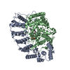
| Title | CRYSTAL STRUCTURE OF CANDIDA ALBICANS PROTEIN FARNESYLTRANSFERASE BINARY COMPLEX WITH THE ISOPRENOID FARNESYLDIPHOSPHATE | ||||||
 Components Components | (Protein farnesyltransferase/geranylgeranyltransferase type-1 ...) x 2 | ||||||
 Keywords Keywords |  TRANSFERASE / Farnesyl transferase TRANSFERASE / Farnesyl transferase | ||||||
| Function / homology |  Function and homology information Function and homology informationProtein prenylyltransferase / Glycosyltransferase - #20 / Glycosyltransferase / Alpha/alpha barrel / Serine Threonine Protein Phosphatase 5, Tetratricopeptide repeat / Alpha Horseshoe / Mainly AlphaSimilarity search - Domain/homology | ||||||
| Biological species |  Candida albicans (yeast) Candida albicans (yeast) | ||||||
| Method | X-RAY DIFFRACTION / SYNCHROTRON / MOLECULAR REPLACEMENT / Resolution: 2.701 Å | ||||||
 Authors Authors | Kumar, S. / Mabanglo, M.F. / Hast, M.A. / Shi, Y. / Beese, L.S. | ||||||
| Funding support |  United States, 1items United States, 1items
| ||||||
 Citation Citation | Journal: To Be Published Title: CRYSTAL STRUCTURE OF CANDIDA ALBICANS PROTEIN FARNESYLTRANSFERASE BINARY COMPLEX WITH THE ISOPRENOID FARNESYLDIPHOSPHATE Authors: Kumar, S. / Mabanglo, M.F. / Hast, M.A. / Shi, Y. / Beese, L.S. | ||||||
| History |
|
- Structure visualization
| Structure viewer | Molecule: MolmilJmol/JSmol |
|---|
- Downloads & links
-Download
| PDBx/mmCIF format | 4yde.cif.gz | 314.3 KB | Display | PDBx/mmCIF format |
|---|---|---|---|---|
| PDB format | pdb4yde.ent.gz | 253.4 KB | Display | PDB format |
| PDBx/mmJSON format | 4yde.json.gz | Tree view | PDBx/mmJSON format | |
| Others |  Other downloads Other downloads |
-Validation report
| Arichive directory | https://data.pdbj.org/pub/pdb/validation_reports/yd/4ydeftp://data.pdbj.org/pub/pdb/validation_reports/yd/4yde | HTTPS FTP |
|---|
-Related structure data
| Related structure data |  4l9pS S: Starting model for refinement |
|---|---|
| Similar structure data |


-Links
 PDBj
PDBj- Assembly
| Deposited unit | 
| ||||||||
|---|---|---|---|---|---|---|---|---|---|
| 1 |
| ||||||||
| Unit cell |
|
-Components
-Protein farnesyltransferase/geranylgeranyltransferase type-1 ... , 2 types, 2 molecules AB
| #1: Protein | Mass: 39236.766 Da / Num. of mol.: 1 Source method: isolated from a genetically manipulated source Source: (gene. exp.) Candida albicans (strain SC5314 / ATCC MYA-2876) (yeast)Strain: SC5314 / ATCC MYA-2876 / Gene: RAM2, CaO19.12280, CaO19.4817 / Production host:  Escherichia coli (E. coli) / References: UniProt: Q5APE8 Escherichia coli (E. coli) / References: UniProt: Q5APE8 |
|---|---|
| #2: Protein | Mass: 66693.289 Da / Num. of mol.: 1 Source method: isolated from a genetically manipulated source Source: (gene. exp.) Candida albicans (strain SC5314 / ATCC MYA-2876) (yeast)Strain: SC5314 / ATCC MYA-2876 / Gene: RAM1, CaO19.12513 / Production host: Escherichia coli (E. coli) / References: UniProt: Q59LE1 |
-Non-polymers , 4 types, 154 molecules 


| #3: Chemical | Ethylene glycol Mass: 62.068 Da / Num. of mol.: 2 / Source method: obtained synthetically / Formula: C2H6O2 Mass: 62.068 Da / Num. of mol.: 2 / Source method: obtained synthetically / Formula: C2H6O2#4: Chemical | ChemComp-ZN / |  Mass: 65.409 Da / Num. of mol.: 1 / Source method: obtained synthetically / Formula: Zn Mass: 65.409 Da / Num. of mol.: 1 / Source method: obtained synthetically / Formula: Zn#5: Chemical | ChemComp-4C7 / ( |  Mass: 388.374 Da / Num. of mol.: 1 / Source method: obtained synthetically / Formula: C15H34O7P2 Mass: 388.374 Da / Num. of mol.: 1 / Source method: obtained synthetically / Formula: C15H34O7P2#6: Water | ChemComp-HOH / | WaterMass: 18.015 Da / Num. of mol.: 150 / Source method: isolated from a natural source / Formula: H2O |
|---|
-Experimental details
-Experiment
| Experiment | Method: X-RAY DIFFRACTION / Number of used crystals: 1 |
|---|
- Sample preparation
| Crystal | Density Matthews: 2.23 Å3/Da / Density % sol: 45 % |
|---|---|
| Crystal grow | Temperature: 290 K / Method: vapor diffusion, sitting drop / pH: 7.2 Details: 9%-13% PEG 2050 chips, 100 mM HEPES pH 7.2 100 -200 mM CaCl2, 5 uM ZnCl2 |
-Data collection
| Diffraction | Mean temperature: 100 K |
|---|---|
| Diffraction source | Source: SYNCHROTRON / Site: APS / Beamline: 22-BM / Wavelength: 1 Å |
| Detector | Type: MARMOSAIC 225 mm CCD / Detector: CCD / Date: Aug 13, 2009 |
| Radiation | Protocol: SINGLE WAVELENGTH / Monochromatic (M) / Laue (L): M / Scattering type: x-ray |
| Radiation wavelength | Wavelength: 1 Å / Relative weight: 1 |
| Reflection | Resolution: 2.7→47.82 Å / Num. obs: 26416 / % possible obs: 100 % / Redundancy: 14.5 % / Rsym value: 0.118 / Net I/σ(I): 31.1 |
| Reflection shell | Resolution: 2.7→2.79 Å / Redundancy: 14.8 % / Rmerge(I) obs: 0.67 / Mean I/σ(I) obs: 4.2 / % possible all: 100 |
- Processing
| Software |
| |||||||||||||||||||||||||||||||||||||||||||||||||||||||||||||||||||||||||||||||||||||||||||||||||||||||||
|---|---|---|---|---|---|---|---|---|---|---|---|---|---|---|---|---|---|---|---|---|---|---|---|---|---|---|---|---|---|---|---|---|---|---|---|---|---|---|---|---|---|---|---|---|---|---|---|---|---|---|---|---|---|---|---|---|---|---|---|---|---|---|---|---|---|---|---|---|---|---|---|---|---|---|---|---|---|---|---|---|---|---|---|---|---|---|---|---|---|---|---|---|---|---|---|---|---|---|---|---|---|---|---|---|---|---|
| Refinement | Method to determine structure: MOLECULAR REPLACEMENT Starting model: 4L9P Resolution: 2.701→47.822 Å / SU ML: 0.32 / Cross valid method: FREE R-VALUE / σ(F): 1.34 / Phase error: 23.49 / Stereochemistry target values: ML
| |||||||||||||||||||||||||||||||||||||||||||||||||||||||||||||||||||||||||||||||||||||||||||||||||||||||||
| Solvent computation | Shrinkage radii: 0.9 Å / VDW probe radii: 1.11 Å / Solvent model: FLAT BULK SOLVENT MODEL | |||||||||||||||||||||||||||||||||||||||||||||||||||||||||||||||||||||||||||||||||||||||||||||||||||||||||
| Refinement step | Cycle: LAST / Resolution: 2.701→47.822 Å
| |||||||||||||||||||||||||||||||||||||||||||||||||||||||||||||||||||||||||||||||||||||||||||||||||||||||||
| Refine LS restraints |
| |||||||||||||||||||||||||||||||||||||||||||||||||||||||||||||||||||||||||||||||||||||||||||||||||||||||||
| LS refinement shell |
| |||||||||||||||||||||||||||||||||||||||||||||||||||||||||||||||||||||||||||||||||||||||||||||||||||||||||
| Refinement TLS params. | Method: refined / Refine-ID: X-RAY DIFFRACTION
| |||||||||||||||||||||||||||||||||||||||||||||||||||||||||||||||||||||||||||||||||||||||||||||||||||||||||
| Refinement TLS group |
|
