 Movie
Movie Controller


+ Open data

- Basic information
| Entry | Database: PDB / ID: 5ohg | ||||||
|---|---|---|---|---|---|---|---|
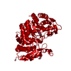
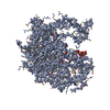
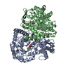
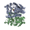
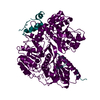
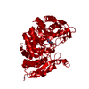
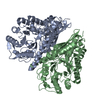
| Title | enolase in complex with RNase E | ||||||
 Components Components |
| ||||||
 Keywords Keywords |  LYASE / enolase / RNase E LYASE / enolase / RNase E | ||||||
| Function / homology |  Function and homology informationregulation of RNA helicase activity / rRNA 5'-end processing / ribonuclease E / ribonuclease E activity / bacterial degradosome / endoribonuclease complex / DEAD/H-box RNA helicase binding / phosphopyruvate hydratase / phosphopyruvate hydratase complex / phosphopyruvate hydratase activity ...regulation of RNA helicase activity / rRNA 5'-end processing / ribonuclease E / ribonuclease E activity / bacterial degradosome / endoribonuclease complex / DEAD/H-box RNA helicase binding / phosphopyruvate hydratase / phosphopyruvate hydratase complex / phosphopyruvate hydratase activity / 7S RNA binding / RNA catabolic process / tRNA processing / mRNA catabolic process / RNA nuclease activity / RNA processing / RNA endonuclease activity / glycolytic process / cytoplasmic side of plasma membrane / rRNA processing / protein complex oligomerization / protein homotetramerization / tRNA binding / rRNA binding / molecular adaptor activity / cytoskeleton / magnesium ion binding / cell surface / protein homodimerization activity / RNA binding / zinc ion binding / extracellular region / membrane / identical protein binding / cytosol / cytoplasm Function and homology informationregulation of RNA helicase activity / rRNA 5'-end processing / ribonuclease E / ribonuclease E activity / bacterial degradosome / endoribonuclease complex / DEAD/H-box RNA helicase binding / phosphopyruvate hydratase / phosphopyruvate hydratase complex / phosphopyruvate hydratase activity ...regulation of RNA helicase activity / rRNA 5'-end processing / ribonuclease E / ribonuclease E activity / bacterial degradosome / endoribonuclease complex / DEAD/H-box RNA helicase binding / phosphopyruvate hydratase / phosphopyruvate hydratase complex / phosphopyruvate hydratase activity / 7S RNA binding / RNA catabolic process / tRNA processing / mRNA catabolic process / RNA nuclease activity / RNA processing / RNA endonuclease activity / glycolytic process / cytoplasmic side of plasma membrane / rRNA processing / protein complex oligomerization / protein homotetramerization / tRNA binding / rRNA binding / molecular adaptor activity / cytoskeleton / magnesium ion binding / cell surface / protein homodimerization activity / RNA binding / zinc ion binding / extracellular region / membrane / identical protein binding / cytosol / cytoplasmSimilarity search - Function | ||||||
| Biological species |  Escherichia coli (E. coli) Escherichia coli (E. coli) | ||||||
| Method | X-RAY DIFFRACTION / SYNCHROTRON / MOLECULAR REPLACEMENT / Resolution: 1.997 Å | ||||||
 Authors Authors | Du, D. / Luisi, B.F. | ||||||
| Funding support |  United Kingdom, 1items United Kingdom, 1items
| ||||||
 Citation Citation | Journal: Nucleic Acids Res / Year: 2018 Title: Analysis of the natively unstructured RNA/protein-recognition core in the Escherichia coli RNA degradosome and its interactions with regulatory RNA/Hfq complexes. Authors: Heather A Bruce / Dijun Du / Dijana Matak-Vinkovic / Katarzyna J Bandyra / R William Broadhurst / Esther Martin / Frank Sobott / Alexander V Shkumatov / Ben F Luisi /  Abstract: The RNA degradosome is a multi-enzyme assembly that plays a central role in the RNA metabolism of Escherichia coli and numerous other bacterial species including pathogens. At the core of the ...The RNA degradosome is a multi-enzyme assembly that plays a central role in the RNA metabolism of Escherichia coli and numerous other bacterial species including pathogens. At the core of the assembly is the endoribonuclease RNase E, one of the largest E. coli proteins and also one that bears the greatest region predicted to be natively unstructured. This extensive unstructured region, situated in the C-terminal half of RNase E, is punctuated with conserved short linear motifs that recruit partner proteins, direct RNA interactions, and enable association with the cytoplasmic membrane. We have structurally characterized a subassembly of the degradosome-comprising a 248-residue segment of the natively unstructured part of RNase E, the DEAD-box helicase RhlB and the glycolytic enzyme enolase, and provide evidence that it serves as a flexible recognition centre that can co-recruit small regulatory RNA and the RNA chaperone Hfq. Our results support a model in which the degradosome captures substrates and regulatory RNAs through the recognition centre, facilitates pairing to cognate transcripts and presents the target to the ribonuclease active sites of the greater assembly for cooperative degradation or processing. | ||||||
| History |
|
- Structure visualization
| Structure viewer | Molecule: MolmilJmol/JSmol |
|---|
- Downloads & links
-Download
| PDBx/mmCIF format | 5ohg.cif.gz | 378.5 KB | Display | PDBx/mmCIF format |
|---|---|---|---|---|
| PDB format | pdb5ohg.ent.gz | 303.3 KB | Display | PDB format |
| PDBx/mmJSON format | 5ohg.json.gz | Tree view | PDBx/mmJSON format | |
| Others |  Other downloads Other downloads |
-Validation report
| Arichive directory | https://data.pdbj.org/pub/pdb/validation_reports/oh/5ohgftp://data.pdbj.org/pub/pdb/validation_reports/oh/5ohg | HTTPS FTP |
|---|
-Related structure data
| Related structure data | 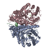 2fymS S: Starting model for refinement C: citing same article ( |
|---|---|
| Similar structure data |


-Links
 PDBj
PDBj


- Assembly
| Deposited unit | 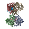
| ||||||||
|---|---|---|---|---|---|---|---|---|---|
| 1 | 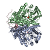
| ||||||||
| 2 | 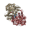
| ||||||||
| Unit cell |
|
-Components
-Protein , 1 types, 4 molecules ABHI
| #1: Protein | / 2-phospho-D-glycerate hydro-lyase / 2-phosphoglycerate dehydratase Mass: 45709.812 Da / Num. of mol.: 4 Source method: isolated from a genetically manipulated source Source: (gene. exp.) Escherichia coli (strain K12) (bacteria)Gene: eno, b2779, JW2750 / Production host: Escherichia coli (E. coli) / References: UniProt: P0A6P9, phosphopyruvate hydratase |
|---|
-Ribonuclease ... , 2 types, 2 molecules CJ
| #2: Protein/peptide | / RNase E Mass: 4214.870 Da / Num. of mol.: 1 Source method: isolated from a genetically manipulated source Details: RNase E Source: (gene. exp.) Escherichia coli (strain K12) (bacteria)Gene: rne, ams, hmp1, b1084, JW1071 / Production host: Escherichia coli (E. coli) / References: UniProt: P21513, ribonuclease E |
|---|---|
| #3: Protein/peptide | / RNase E Mass: 3993.634 Da / Num. of mol.: 1 Source method: isolated from a genetically manipulated source Source: (gene. exp.) Escherichia coli (strain K12) (bacteria)Gene: rne, ams, hmp1, b1084, JW1071 / Production host: Escherichia coli (E. coli) / References: UniProt: P21513, ribonuclease E |
-Non-polymers , 4 types, 1689 molecules 


| #4: Chemical | ChemComp-MG /  Mass: 24.305 Da / Num. of mol.: 4 / Source method: obtained synthetically / Formula: Mg Mass: 24.305 Da / Num. of mol.: 4 / Source method: obtained synthetically / Formula: Mg#5: Chemical | ChemComp-NA /  Mass: 22.990 Da / Num. of mol.: 5 / Source method: obtained synthetically / Formula: Na Mass: 22.990 Da / Num. of mol.: 5 / Source method: obtained synthetically / Formula: Na#6: Chemical | ChemComp-PO4 / Phosphate Mass: 94.971 Da / Num. of mol.: 4 / Source method: obtained synthetically / Formula: PO4 Mass: 94.971 Da / Num. of mol.: 4 / Source method: obtained synthetically / Formula: PO4#7: Water | ChemComp-HOH / | WaterMass: 18.015 Da / Num. of mol.: 1676 / Source method: isolated from a natural source / Formula: H2O |
|---|
-Experimental details
-Experiment
| Experiment | Method: X-RAY DIFFRACTION / Number of used crystals: 1 |
|---|
- Sample preparation
| Crystal | Density Matthews: 2.37 Å3/Da / Density % sol: 48.19 % |
|---|---|
| Crystal grow | Temperature: 293 K / Method: vapor diffusion, sitting drop / pH: 8.5 Details: 0.2 M potassium thiocyanate, 0.1 M Bis-Tris propane (pH 8.5) and 20% (w/v) PEG 3350 |
-Data collection
| Diffraction | Mean temperature: 100 K |
|---|---|
| Diffraction source | Source: SYNCHROTRON / Site: Diamond / Beamline: I04 / Wavelength: 1 Å |
| Detector | Type: DECTRIS PILATUS 300K / Detector: PIXEL / Date: Nov 1, 2009 |
| Radiation | Protocol: SINGLE WAVELENGTH / Monochromatic (M) / Laue (L): M / Scattering type: x-ray |
| Radiation wavelength | Wavelength: 1 Å / Relative weight: 1 |
| Reflection | Resolution: 1.997→40 Å / Num. all: 121616 / Num. obs: 118999 / % possible obs: 98.59 % / Redundancy: 3.7 % / Rmerge(I) obs: 0.099 / Rpim(I) all: 0.061 / Net I/σ(I): 9.2 |
| Reflection shell | Resolution: 1.99→2.09 Å / Rmerge(I) obs: 0.301 / Rpim(I) all: 0.191 |
- Processing
| Software |
| |||||||||||||||||||||||||||||||||||||||||||||||||||||||||||||||||||||||||||||||||||||||||||||||||||||||||||||||||||||||||||||||||||||||||||||||||||||||||||||||||||||||||||||||||||||||||||||||||||||||||||||||||||||||||
|---|---|---|---|---|---|---|---|---|---|---|---|---|---|---|---|---|---|---|---|---|---|---|---|---|---|---|---|---|---|---|---|---|---|---|---|---|---|---|---|---|---|---|---|---|---|---|---|---|---|---|---|---|---|---|---|---|---|---|---|---|---|---|---|---|---|---|---|---|---|---|---|---|---|---|---|---|---|---|---|---|---|---|---|---|---|---|---|---|---|---|---|---|---|---|---|---|---|---|---|---|---|---|---|---|---|---|---|---|---|---|---|---|---|---|---|---|---|---|---|---|---|---|---|---|---|---|---|---|---|---|---|---|---|---|---|---|---|---|---|---|---|---|---|---|---|---|---|---|---|---|---|---|---|---|---|---|---|---|---|---|---|---|---|---|---|---|---|---|---|---|---|---|---|---|---|---|---|---|---|---|---|---|---|---|---|---|---|---|---|---|---|---|---|---|---|---|---|---|---|---|---|---|---|---|---|---|---|---|---|---|---|---|---|---|---|---|---|---|
| Refinement | Method to determine structure: MOLECULAR REPLACEMENT Starting model: 2fym Resolution: 1.997→39.244 Å / SU ML: 0.19 / Cross valid method: FREE R-VALUE / σ(F): 1.33 / Phase error: 19.07
| |||||||||||||||||||||||||||||||||||||||||||||||||||||||||||||||||||||||||||||||||||||||||||||||||||||||||||||||||||||||||||||||||||||||||||||||||||||||||||||||||||||||||||||||||||||||||||||||||||||||||||||||||||||||||
| Solvent computation | Shrinkage radii: 0.9 Å / VDW probe radii: 1.11 Å | |||||||||||||||||||||||||||||||||||||||||||||||||||||||||||||||||||||||||||||||||||||||||||||||||||||||||||||||||||||||||||||||||||||||||||||||||||||||||||||||||||||||||||||||||||||||||||||||||||||||||||||||||||||||||
| Refinement step | Cycle: LAST / Resolution: 1.997→39.244 Å
| |||||||||||||||||||||||||||||||||||||||||||||||||||||||||||||||||||||||||||||||||||||||||||||||||||||||||||||||||||||||||||||||||||||||||||||||||||||||||||||||||||||||||||||||||||||||||||||||||||||||||||||||||||||||||
| Refine LS restraints |
| |||||||||||||||||||||||||||||||||||||||||||||||||||||||||||||||||||||||||||||||||||||||||||||||||||||||||||||||||||||||||||||||||||||||||||||||||||||||||||||||||||||||||||||||||||||||||||||||||||||||||||||||||||||||||
| LS refinement shell |
|
