 Movie
Movie Controller

[English] 日本語
 Yorodumi
- PDB-4l9p: Crystal structure of Aspergillus fumigatus protein farnesyltransf... -
+ Open data

- Basic information
| Entry | Database: PDB / ID: 4l9p | ||||||
|---|---|---|---|---|---|---|---|
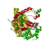
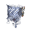
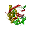
| Title | Crystal structure of Aspergillus fumigatus protein farnesyltransferase complexed with the FII analog, FPT-II, and the KCVVM peptide | ||||||
 Components Components |
| ||||||
 Keywords Keywords |  TRANSFERASE / Ternary complex with isoprenoid and CAAX peptide substrate / CAAX farnesyltransferase / Isoprenoid and CAAX protein/peptide substrate TRANSFERASE / Ternary complex with isoprenoid and CAAX peptide substrate / CAAX farnesyltransferase / Isoprenoid and CAAX protein/peptide substrate | ||||||
| Function / homology |  Function and homology information Function and homology informationpeptide pheromone maturation / prenylation / CAAX-protein geranylgeranyltransferase activity / CAAX-protein geranylgeranyltransferase complex / protein geranylgeranyltransferase type I / protein farnesyltransferase / protein farnesyltransferase activity / protein farnesyltransferase complex / zinc ion binding / cytoplasmSimilarity search - Function | ||||||
| Biological species |  Aspergillus fumigatus (mold) Aspergillus fumigatus (mold) | ||||||
| Method | X-RAY DIFFRACTION / SYNCHROTRON / MOLECULAR REPLACEMENT / Resolution: 1.45 Å | ||||||
 Authors Authors | Mabanglo, M.F. / Hast, M.A. / Beese, L.S. | ||||||
 Citation Citation | Journal: Protein Sci. / Year: 2014 Title: Crystal structures of the fungal pathogen Aspergillus fumigatus protein farnesyltransferase complexed with substrates and inhibitors reveal features for antifungal drug design. Authors: Mabanglo, M.F. / Hast, M.A. / Lubock, N.B. / Hellinga, H.W. / Beese, L.S. | ||||||
| History |
|
- Structure visualization
| Structure viewer | Molecule: MolmilJmol/JSmol |
|---|
- Downloads & links
-Download
| PDBx/mmCIF format | 4l9p.cif.gz | 372.4 KB | Display | PDBx/mmCIF format |
|---|---|---|---|---|
| PDB format | pdb4l9p.ent.gz | 297.5 KB | Display | PDB format |
| PDBx/mmJSON format | 4l9p.json.gz | Tree view | PDBx/mmJSON format | |
| Others |  Other downloads Other downloads |
-Validation report
| Arichive directory | https://data.pdbj.org/pub/pdb/validation_reports/l9/4l9pftp://data.pdbj.org/pub/pdb/validation_reports/l9/4l9p | HTTPS FTP |
|---|
-Related structure data


-Links
 PDBj
PDBj

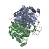
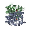
- Assembly
| Deposited unit | 
| ||||||||
|---|---|---|---|---|---|---|---|---|---|
| 1 |
| ||||||||
| Unit cell |
|
-Components
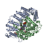
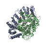
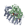
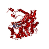
-CaaX farnesyltransferase ... , 2 types, 2 molecules AB
| #1: Protein | Mass: 42391.461 Da / Num. of mol.: 1 / Mutation: N146S Source method: isolated from a genetically manipulated source Source: (gene. exp.) Aspergillus fumigatus (mold) / Strain: Af293 / Gene: AFUA_4G07800 / Plasmid: pCDF-Duet1 / Production host:  Escherichia coli (E. coli) / Strain (production host): C41(DE3) Escherichia coli (E. coli) / Strain (production host): C41(DE3)References: UniProt: Q4WP27, Transferases; Transferring alkyl or aryl groups, other than methyl groups |
|---|---|
| #2: Protein | Mass: 56635.805 Da / Num. of mol.: 1 Source method: isolated from a genetically manipulated source Source: (gene. exp.) Aspergillus fumigatus (mold) / Strain: Af293 / Gene: AFUA_4G10330 / Plasmid: pCDF-Duet1 / Production host: Escherichia coli (E. coli) / Strain (production host): C41(DE3) / References: UniProt: Q4WPS9, protein farnesyltransferase |
-Protein/peptide , 1 types, 1 molecules C
| #3: Protein/peptide | Mass: 579.795 Da / Num. of mol.: 1 / Source method: obtained synthetically |
|---|
-Non-polymers , 5 types, 792 molecules 


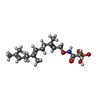

| #4: Chemical | Chloride Mass: 35.453 Da / Num. of mol.: 3 / Source method: obtained synthetically / Formula: Cl Mass: 35.453 Da / Num. of mol.: 3 / Source method: obtained synthetically / Formula: Cl#5: Chemical | ChemComp-EDO / Ethylene glycol Mass: 62.068 Da / Num. of mol.: 9 / Source method: obtained synthetically / Formula: C2H6O2 Mass: 62.068 Da / Num. of mol.: 9 / Source method: obtained synthetically / Formula: C2H6O2#6: Chemical | ChemComp-ZN / |  Mass: 65.409 Da / Num. of mol.: 1 / Source method: obtained synthetically / Formula: Zn Mass: 65.409 Da / Num. of mol.: 1 / Source method: obtained synthetically / Formula: Zn#7: Chemical | ChemComp-FII / [( |  Mass: 359.398 Da / Num. of mol.: 1 / Source method: obtained synthetically / Formula: C17H30NO5P Mass: 359.398 Da / Num. of mol.: 1 / Source method: obtained synthetically / Formula: C17H30NO5P#8: Water | ChemComp-HOH / | WaterMass: 18.015 Da / Num. of mol.: 778 / Source method: isolated from a natural source / Formula: H2O |
|---|
-Experimental details
-Experiment
| Experiment | Method: X-RAY DIFFRACTION / Number of used crystals: 1 |
|---|
- Sample preparation
| Crystal | Density Matthews: 2.22 Å3/Da / Density % sol: 44.64 % |
|---|---|
| Crystal grow | Temperature: 290 K / Method: vapor diffusion, hanging drop / pH: 7.5 Details: 4-10% PEG6000, 600-800 mM LiCl, 100 mM HEPES, pH 7.5, VAPOR DIFFUSION, HANGING DROP, temperature 290K |
-Data collection
| Diffraction | Mean temperature: 100 K |
|---|---|
| Diffraction source | Source: SYNCHROTRON / Site: APS  / Beamline: 22-BM / Wavelength: 1 Å / Beamline: 22-BM / Wavelength: 1 Å |
| Detector | Type: MARMOSAIC 225 mm CCD / Detector: CCD / Date: Jun 8, 2012 |
| Radiation | Monochromator: Double-crystal, liquid nitrogen cooled / Protocol: SINGLE WAVELENGTH / Monochromatic (M) / Laue (L): M / Scattering type: x-ray |
| Radiation wavelength | Wavelength: 1 Å / Relative weight: 1 |
| Reflection | Resolution: 1.45→50 Å / Num. all: 156214 / Num. obs: 154808 / % possible obs: 99.1 % / Observed criterion σ(F): 1 / Observed criterion σ(I): 1 / Rmerge(I) obs: 0.069 / Net I/σ(I): 36 |
| Reflection shell | Resolution: 1.45→1.48 Å / Redundancy: 5.5 % / Rmerge(I) obs: 0.591 / Mean I/σ(I) obs: 2 / % possible all: 82.8 |
- Processing
| Software |
| |||||||||||||||||||||||||||||||||||||||||||||||||||||||||||||||||||||||||||||||||||||||||||||||||||||||||||||||||||||||||||||||||||||||||||||||||||||||||||||||||||||||||||||||||||||||||||||||||||||||||||||||||||||||||
|---|---|---|---|---|---|---|---|---|---|---|---|---|---|---|---|---|---|---|---|---|---|---|---|---|---|---|---|---|---|---|---|---|---|---|---|---|---|---|---|---|---|---|---|---|---|---|---|---|---|---|---|---|---|---|---|---|---|---|---|---|---|---|---|---|---|---|---|---|---|---|---|---|---|---|---|---|---|---|---|---|---|---|---|---|---|---|---|---|---|---|---|---|---|---|---|---|---|---|---|---|---|---|---|---|---|---|---|---|---|---|---|---|---|---|---|---|---|---|---|---|---|---|---|---|---|---|---|---|---|---|---|---|---|---|---|---|---|---|---|---|---|---|---|---|---|---|---|---|---|---|---|---|---|---|---|---|---|---|---|---|---|---|---|---|---|---|---|---|---|---|---|---|---|---|---|---|---|---|---|---|---|---|---|---|---|---|---|---|---|---|---|---|---|---|---|---|---|---|---|---|---|---|---|---|---|---|---|---|---|---|---|---|---|---|---|---|---|---|
| Refinement | Method to determine structure: MOLECULAR REPLACEMENT Starting model: > Homology model of A. fumigatus farnesyltransferase generated using PHYRE Resolution: 1.45→22.421 Å / SU ML: 0.12 / σ(F): 1.33 / Phase error: 13.68 / Stereochemistry target values: ML
| |||||||||||||||||||||||||||||||||||||||||||||||||||||||||||||||||||||||||||||||||||||||||||||||||||||||||||||||||||||||||||||||||||||||||||||||||||||||||||||||||||||||||||||||||||||||||||||||||||||||||||||||||||||||||
| Solvent computation | Shrinkage radii: 0.9 Å / VDW probe radii: 1.11 Å / Solvent model: FLAT BULK SOLVENT MODEL | |||||||||||||||||||||||||||||||||||||||||||||||||||||||||||||||||||||||||||||||||||||||||||||||||||||||||||||||||||||||||||||||||||||||||||||||||||||||||||||||||||||||||||||||||||||||||||||||||||||||||||||||||||||||||
| Refinement step | Cycle: LAST / Resolution: 1.45→22.421 Å
| |||||||||||||||||||||||||||||||||||||||||||||||||||||||||||||||||||||||||||||||||||||||||||||||||||||||||||||||||||||||||||||||||||||||||||||||||||||||||||||||||||||||||||||||||||||||||||||||||||||||||||||||||||||||||
| Refine LS restraints |
| |||||||||||||||||||||||||||||||||||||||||||||||||||||||||||||||||||||||||||||||||||||||||||||||||||||||||||||||||||||||||||||||||||||||||||||||||||||||||||||||||||||||||||||||||||||||||||||||||||||||||||||||||||||||||
| LS refinement shell |
|
