 Movie
Movie Controller

[English] 日本語
 Yorodumi
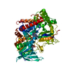
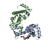
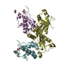
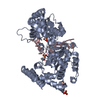
- PDB-4yc7: Crystal structure of human FMNL2 GBD-FH3 Domains bound to Cdc42-GppNHp -
+ Open data

- Basic information
| Entry | Database: PDB / ID: 4yc7 | ||||||
|---|---|---|---|---|---|---|---|
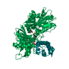
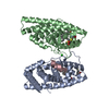
| Title | Crystal structure of human FMNL2 GBD-FH3 Domains bound to Cdc42-GppNHp | ||||||
 Components Components |
| ||||||
 Keywords Keywords |  SIGNALING PROTEIN / Armadillo repeat / Rho GTPase / cell cycle SIGNALING PROTEIN / Armadillo repeat / Rho GTPase / cell cycle | ||||||
| Function / homology |  Function and homology information Function and homology informationsmall GTPase binding => GO:0031267 / GBD domain binding / submandibular salivary gland formation / actin filament branching / Golgi transport complex / positive regulation of pinocytosis / modification of synaptic structure / endothelin receptor signaling pathway involved in heart process / Cdc42 protein signal transduction / cardiac neural crest cell migration involved in outflow tract morphogenesis ...small GTPase binding => GO:0031267 / GBD domain binding / submandibular salivary gland formation / actin filament branching / Golgi transport complex / positive regulation of pinocytosis / modification of synaptic structure / endothelin receptor signaling pathway involved in heart process / Cdc42 protein signal transduction / cardiac neural crest cell migration involved in outflow tract morphogenesis / positive regulation of synapse structural plasticity / dendritic cell migration / storage vacuole / positive regulation of epithelial cell proliferation involved in lung morphogenesis / apolipoprotein A-I receptor binding / neuron fate determination / modulation by host of viral process / GTP-dependent protein binding / organelle transport along microtubule / regulation of attachment of spindle microtubules to kinetochore / positive regulation of pseudopodium assembly / Inactivation of CDC42 and RAC1 / cardiac conduction system development / regulation of filopodium assembly / establishment of Golgi localization / leading edge membrane / neuropilin signaling pathway / positive regulation of intracellular protein transport / cell junction assembly / filopodium assembly / establishment of epithelial cell apical/basal polarity / regulation of modification of postsynaptic structure / mitogen-activated protein kinase kinase kinase binding / dendritic spine morphogenesis / thioesterase binding / embryonic heart tube development / regulation of stress fiber assembly / regulation of cell morphogenesis / RHO GTPases activate KTN1 / regulation of lamellipodium assembly / nuclear migration / DCC mediated attractive signaling / adherens junction organization / sprouting angiogenesis / Wnt signaling pathway, planar cell polarity pathway / CD28 dependent Vav1 pathway / regulation of postsynapse organization / positive regulation of filopodium assembly / cortical actin cytoskeleton organization / regulation of mitotic nuclear division / phagocytosis, engulfment / RHOV GTPase cycle / establishment or maintenance of cell polarity / heart contraction / Myogenesis / RHOJ GTPase cycle / Golgi organization / RHOQ GTPase cycle / positive regulation of cytokinesis / RHO GTPases activate PAKs / CDC42 GTPase cycle / RHOU GTPase cycle / macrophage differentiation / RHOG GTPase cycle / RHO GTPases Activate WASPs and WAVEs / RAC2 GTPase cycle / RHO GTPases activate IQGAPs / RAC3 GTPase cycle / spindle midzone / negative regulation of protein-containing complex assembly / positive regulation of lamellipodium assembly / positive regulation of substrate adhesion-dependent cell spreading / phagocytic vesicle / positive regulation of stress fiber assembly / GPVI-mediated activation cascade / cytoskeleton organization / RAC1 GTPase cycle / EPHB-mediated forward signaling / substantia nigra development / Gene and protein expression by JAK-STAT signaling after Interleukin-12 stimulation / small monomeric GTPase / G protein activity / secretory granule / positive regulation of DNA replication / filopodium / integrin-mediated signaling pathway / RHO GTPases Activate Formins / actin filament organization / regulation of actin cytoskeleton organization / FCGR3A-mediated phagocytosis / EGFR downregulation / positive regulation of JNK cascade / MAPK6/MAPK4 signaling / Schaffer collateral - CA1 synapse / protein localization / G beta:gamma signalling through CDC42 / mitotic spindle / Regulation of actin dynamics for phagocytic cup formation / VEGFA-VEGFR2 Pathway / cellular response to type II interferonSimilarity search - Function | ||||||
| Biological species |  Homo sapiens (human) Homo sapiens (human) | ||||||
| Method | X-RAY DIFFRACTION / SYNCHROTRON / MOLECULAR REPLACEMENT / Resolution: 2.5 Å | ||||||
 Authors Authors | Kuhn, S. / Geyer, M. | ||||||
 Citation Citation | Journal: Nat Commun / Year: 2015 Title: The structure of FMNL2-Cdc42 yields insights into the mechanism of lamellipodia and filopodia formation. Authors: Kuhn, S. / Erdmann, C. / Kage, F. / Block, J. / Schwenkmezger, L. / Steffen, A. / Rottner, K. / Geyer, M. | ||||||
| History |
|
- Structure visualization
| Structure viewer | Molecule: MolmilJmol/JSmol |
|---|
- Downloads & links
-Download
| PDBx/mmCIF format | 4yc7.cif.gz | 211.5 KB | Display | PDBx/mmCIF format |
|---|---|---|---|---|
| PDB format | pdb4yc7.ent.gz | 167.3 KB | Display | PDB format |
| PDBx/mmJSON format | 4yc7.json.gz | Tree view | PDBx/mmJSON format | |
| Others |  Other downloads Other downloads |
-Validation report
| Arichive directory | https://data.pdbj.org/pub/pdb/validation_reports/yc/4yc7ftp://data.pdbj.org/pub/pdb/validation_reports/yc/4yc7 | HTTPS FTP |
|---|
-Related structure data
| Related structure data | 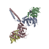 4ydhC 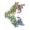 3eg5S S: Starting model for refinement C: citing same article ( |
|---|---|
| Similar structure data |


-Links
 PDBj
PDBj


- Assembly
| Deposited unit | 
| ||||||||
|---|---|---|---|---|---|---|---|---|---|
| 1 |
| ||||||||
| Unit cell |
|
-Components
| #1: Protein | Mass: 44081.191 Da / Num. of mol.: 1 / Fragment: UNP residues 1-379 Source method: isolated from a genetically manipulated source Source: (gene. exp.) Homo sapiens (human) / Gene: FMNL2, FHOD2, KIAA1902 / Production host:  Escherichia coli (E. coli) / References: UniProt: Q96PY5 Escherichia coli (E. coli) / References: UniProt: Q96PY5 |
|---|---|
| #2: Protein | Mass: 20027.961 Da / Num. of mol.: 1 / Fragment: UNP residues 1-179 Source method: isolated from a genetically manipulated source Source: (gene. exp.) Homo sapiens (human) / Gene: CDC42 / Production host: Escherichia coli (E. coli) / References: UniProt: P60953 |
| #3: Chemical | ChemComp-GNP / 5'-Guanylyl imidodiphosphate  Mass: 522.196 Da / Num. of mol.: 1 Mass: 522.196 Da / Num. of mol.: 1Source method: isolated from a genetically manipulated source Formula: C10H17N6O13P3 Comment: GppNHp, GMPPNP, energy-carrying molecule analogue*YM |
| #4: Chemical | ChemComp-MG /   Mass: 24.305 Da / Num. of mol.: 1 Mass: 24.305 Da / Num. of mol.: 1Source method: isolated from a genetically manipulated source Formula: Mg |
| #5: Water | ChemComp-HOH / Water Mass: 18.015 Da / Num. of mol.: 63 / Source method: isolated from a natural source / Formula: H2O Mass: 18.015 Da / Num. of mol.: 63 / Source method: isolated from a natural source / Formula: H2O |
-Experimental details
-Experiment
| Experiment | Method: X-RAY DIFFRACTION |
|---|
- Sample preparation
| Crystal | Density Matthews: 2.33 Å3/Da / Density % sol: 47.2 % |
|---|---|
| Crystal grow | Temperature: 293 K / Method: vapor diffusion, hanging drop / pH: 8 Details: 20 mM Tris/HCl (pH 8.0), 14% (v/v) PEG 3350, 0.25 M magnesium acetate |
-Data collection
| Diffraction | Mean temperature: 100 K |
|---|---|
| Diffraction source | Source: SYNCHROTRON / Site: SLS  / Beamline: X10SA / Wavelength: 1 Å / Beamline: X10SA / Wavelength: 1 Å |
| Detector | Type: MARRESEARCH / Detector: CCD / Date: Jul 20, 2010 |
| Radiation | Protocol: SINGLE WAVELENGTH / Monochromatic (M) / Laue (L): M / Scattering type: x-ray |
| Radiation wavelength | Wavelength: 1 Å / Relative weight: 1 |
| Reflection | Resolution: 2.5→43.4 Å / Num. obs: 22213 / % possible obs: 100 % / Redundancy: 14.1 % / Rmerge(I) obs: 0.053 / Net I/σ(I): 25.9 |
| Reflection shell | Highest resolution: 2.5 Å / Redundancy: 14.7 % / Rmerge(I) obs: 0.336 / Mean I/σ(I) obs: 4.4 / Num. measured obs: 4521 / % possible all: 100 |
- Processing
| Software |
| |||||||||||||||||||||||||||||||||||||||||||||||||||||||||||||||||||||||||||||||||||||||||||||||||||||||||||||||||||||||||||||||||||||||||||||||||||||||||||||||||||||||||||||||||||||||||||||||||||||||||||||||||||||||||||||||||||||||||||||||||||||||||||||||||||||||||||||||||||||||||||||||||||||||||||||||||||||||||||||||||||||
|---|---|---|---|---|---|---|---|---|---|---|---|---|---|---|---|---|---|---|---|---|---|---|---|---|---|---|---|---|---|---|---|---|---|---|---|---|---|---|---|---|---|---|---|---|---|---|---|---|---|---|---|---|---|---|---|---|---|---|---|---|---|---|---|---|---|---|---|---|---|---|---|---|---|---|---|---|---|---|---|---|---|---|---|---|---|---|---|---|---|---|---|---|---|---|---|---|---|---|---|---|---|---|---|---|---|---|---|---|---|---|---|---|---|---|---|---|---|---|---|---|---|---|---|---|---|---|---|---|---|---|---|---|---|---|---|---|---|---|---|---|---|---|---|---|---|---|---|---|---|---|---|---|---|---|---|---|---|---|---|---|---|---|---|---|---|---|---|---|---|---|---|---|---|---|---|---|---|---|---|---|---|---|---|---|---|---|---|---|---|---|---|---|---|---|---|---|---|---|---|---|---|---|---|---|---|---|---|---|---|---|---|---|---|---|---|---|---|---|---|---|---|---|---|---|---|---|---|---|---|---|---|---|---|---|---|---|---|---|---|---|---|---|---|---|---|---|---|---|---|---|---|---|---|---|---|---|---|---|---|---|---|---|---|---|---|---|---|---|---|---|---|---|---|---|---|---|---|---|---|---|---|---|---|---|---|---|---|---|---|---|---|---|---|---|---|---|---|---|---|---|---|---|---|---|---|---|---|---|---|---|---|---|---|---|---|---|---|---|---|---|---|---|---|---|---|---|
| Refinement | Method to determine structure: MOLECULAR REPLACEMENT Starting model: PDB accession code 3EG5, Chain A Resolution: 2.5→43.4 Å / SU ML: 0.48 / Cross valid method: FREE R-VALUE / σ(F): 2.02 / Phase error: 22.09 / Stereochemistry target values: ML
| |||||||||||||||||||||||||||||||||||||||||||||||||||||||||||||||||||||||||||||||||||||||||||||||||||||||||||||||||||||||||||||||||||||||||||||||||||||||||||||||||||||||||||||||||||||||||||||||||||||||||||||||||||||||||||||||||||||||||||||||||||||||||||||||||||||||||||||||||||||||||||||||||||||||||||||||||||||||||||||||||||||
| Solvent computation | Shrinkage radii: 0.98 Å / VDW probe radii: 1.2 Å / Solvent model: FLAT BULK SOLVENT MODEL / Bsol: 52.781 Å2 / ksol: 0.338 e/Å3 | |||||||||||||||||||||||||||||||||||||||||||||||||||||||||||||||||||||||||||||||||||||||||||||||||||||||||||||||||||||||||||||||||||||||||||||||||||||||||||||||||||||||||||||||||||||||||||||||||||||||||||||||||||||||||||||||||||||||||||||||||||||||||||||||||||||||||||||||||||||||||||||||||||||||||||||||||||||||||||||||||||||
| Displacement parameters |
| |||||||||||||||||||||||||||||||||||||||||||||||||||||||||||||||||||||||||||||||||||||||||||||||||||||||||||||||||||||||||||||||||||||||||||||||||||||||||||||||||||||||||||||||||||||||||||||||||||||||||||||||||||||||||||||||||||||||||||||||||||||||||||||||||||||||||||||||||||||||||||||||||||||||||||||||||||||||||||||||||||||
| Refinement step | Cycle: LAST / Resolution: 2.5→43.4 Å
| |||||||||||||||||||||||||||||||||||||||||||||||||||||||||||||||||||||||||||||||||||||||||||||||||||||||||||||||||||||||||||||||||||||||||||||||||||||||||||||||||||||||||||||||||||||||||||||||||||||||||||||||||||||||||||||||||||||||||||||||||||||||||||||||||||||||||||||||||||||||||||||||||||||||||||||||||||||||||||||||||||||
| Refine LS restraints |
| |||||||||||||||||||||||||||||||||||||||||||||||||||||||||||||||||||||||||||||||||||||||||||||||||||||||||||||||||||||||||||||||||||||||||||||||||||||||||||||||||||||||||||||||||||||||||||||||||||||||||||||||||||||||||||||||||||||||||||||||||||||||||||||||||||||||||||||||||||||||||||||||||||||||||||||||||||||||||||||||||||||
| LS refinement shell |
| |||||||||||||||||||||||||||||||||||||||||||||||||||||||||||||||||||||||||||||||||||||||||||||||||||||||||||||||||||||||||||||||||||||||||||||||||||||||||||||||||||||||||||||||||||||||||||||||||||||||||||||||||||||||||||||||||||||||||||||||||||||||||||||||||||||||||||||||||||||||||||||||||||||||||||||||||||||||||||||||||||||
| Refinement TLS params. | Method: refined / Refine-ID: X-RAY DIFFRACTION
| |||||||||||||||||||||||||||||||||||||||||||||||||||||||||||||||||||||||||||||||||||||||||||||||||||||||||||||||||||||||||||||||||||||||||||||||||||||||||||||||||||||||||||||||||||||||||||||||||||||||||||||||||||||||||||||||||||||||||||||||||||||||||||||||||||||||||||||||||||||||||||||||||||||||||||||||||||||||||||||||||||||
| Refinement TLS group |
|
