 Movie
Movie Controller


+ Open data

- Basic information
| Entry | Database: PDB / ID: 4.0E+44 | ||||||
|---|---|---|---|---|---|---|---|
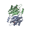
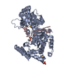
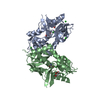
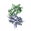
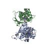
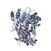
| Title | Crystal structure of the hMHF1/hMHF2 Histone-Fold Tetramer | ||||||
 Components Components |
| ||||||
 Keywords Keywords |  DNA BINDING PROTEIN / Histone-Fold / Tetramer / Fanconi Anemia / FANCM / MHF DNA BINDING PROTEIN / Histone-Fold / Tetramer / Fanconi Anemia / FANCM / MHF | ||||||
| Function / homology |  Function and homology information Function and homology informationFANCM-MHF complex / Fanconi anaemia nuclear complex / resolution of meiotic recombination intermediates / kinetochore assembly / inner kinetochore / replication fork processing / Amplification of signal from unattached kinetochores via a MAD2 inhibitory signal / interstrand cross-link repair / Mitotic Prometaphase / EML4 and NUDC in mitotic spindle formation ...FANCM-MHF complex / Fanconi anaemia nuclear complex / resolution of meiotic recombination intermediates / kinetochore assembly / inner kinetochore / replication fork processing / Amplification of signal from unattached kinetochores via a MAD2 inhibitory signal / interstrand cross-link repair / Mitotic Prometaphase / EML4 and NUDC in mitotic spindle formation / Deposition of new CENPA-containing nucleosomes at the centromere / Resolution of Sister Chromatid Cohesion / positive regulation of protein ubiquitination / chromosome segregation / RHO GTPases Activate Formins / Fanconi Anemia Pathway / PKR-mediated signaling / Separation of Sister Chromatids / protein heterodimerization activity / cell division / DNA repair / DNA damage response / chromatin binding / chromatin / DNA binding / nucleoplasm / nucleus / cytosolSimilarity search - Function | ||||||
| Biological species |  Homo sapiens (human) Homo sapiens (human) | ||||||
| Method | X-RAY DIFFRACTION / SYNCHROTRON / MOLECULAR REPLACEMENT / molecular replacement / Resolution: 2.1 Å | ||||||
 Authors Authors | Fox III, D. / Zhao, Y. / Yang, W. / Weidong, W. | ||||||
 Citation Citation | Journal: To be Published Title: Crystal Structures Reveal that FANCM remodels the MHF Tetramer in favor of binding Branched DNA Authors: Fox III, D. / Yan, Z. / Ling, C. / Zhao, Y. / Lee, D.Y. / Yang, W. / Weidong, W. | ||||||
| History |
|
- Structure visualization
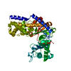
| Structure viewer | Molecule: MolmilJmol/JSmol |
|---|
- Downloads & links
-Download
| PDBx/mmCIF format | 4e44.cif.gz | 82.3 KB | Display | PDBx/mmCIF format |
|---|---|---|---|---|
| PDB format | pdb4e44.ent.gz | 60.8 KB | Display | PDB format |
| PDBx/mmJSON format | 4e44.json.gz | Tree view | PDBx/mmJSON format | |
| Others |  Other downloads Other downloads |
-Validation report
| Arichive directory | https://data.pdbj.org/pub/pdb/validation_reports/e4/4e44ftp://data.pdbj.org/pub/pdb/validation_reports/e4/4e44 | HTTPS FTP |
|---|
-Related structure data
| Related structure data | 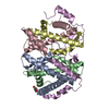 4e45C 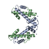 1tafS C: citing same article ( S: Starting model for refinement |
|---|---|
| Similar structure data |


-Links
 PDBj
PDBj


- Assembly
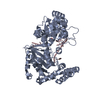
| Deposited unit | 
| ||||||||
|---|---|---|---|---|---|---|---|---|---|
| 1 |
| ||||||||
| Unit cell |
|
-Components
| #1: Protein | / CENP-S / Apoptosis-inducing TAF9-like domain-containing protein 1 / FANCM-interacting histone fold ...CENP-S / Apoptosis-inducing TAF9-like domain-containing protein 1 / FANCM-interacting histone fold protein 1 / Fanconi anemia-associated polypeptide of 16 kDa Mass: 13042.696 Da / Num. of mol.: 2 / Fragment: UNP residues 1-110 Source method: isolated from a genetically manipulated source Source: (gene. exp.) Homo sapiens (human) / Gene: APITD1, CENPS, FAAP16, MHF1 / Plasmid: pGEX/pHis bicistronic / Production host:  Escherichia coli (E. coli) / Strain (production host): BL21(DE3) / References: UniProt: Q8N2Z9 Escherichia coli (E. coli) / Strain (production host): BL21(DE3) / References: UniProt: Q8N2Z9#2: Protein | / CENP-X / FANCM-interacting histone fold protein 2 / Fanconi anemia-associated polypeptide of 10 kDa ...CENP-X / FANCM-interacting histone fold protein 2 / Fanconi anemia-associated polypeptide of 10 kDa / Retinoic acid-inducible gene D9 protein homolog / Stimulated by retinoic acid gene 13 protein homologMass: 9116.545 Da / Num. of mol.: 2 Source method: isolated from a genetically manipulated source Source: (gene. exp.) Homo sapiens (human) / Gene: STRA13, CENPX, FAAP10, MHF2 / Plasmid: pGEX/pHis bicistronic / Production host: Escherichia coli (E. coli) / Strain (production host): BL21(DE3) / References: UniProt: A8MT69#3: Water | ChemComp-HOH / | Water Mass: 18.015 Da / Num. of mol.: 97 / Source method: isolated from a natural source / Formula: H2O Mass: 18.015 Da / Num. of mol.: 97 / Source method: isolated from a natural source / Formula: H2O |
|---|
-Experimental details
-Experiment
| Experiment | Method: X-RAY DIFFRACTION / Number of used crystals: 1 |
|---|
- Sample preparation
| Crystal | Density Matthews: 2.58 Å3/Da / Density % sol: 52.37 % |
|---|---|
| Crystal grow | Temperature: 298 K / Method: vapor diffusion, sitting drop / pH: 7.8 Details: Tracking Code C4, 0.1M Tris pH 7.8, 0.2M LiCl, 0.1M Na2SO4, 17.5% w/v PEG3350, 10% glycerol cryo., vapor diffusion, sitting drop, temperature 298K |
-Data collection
| Diffraction | Mean temperature: 100 K | |||||||||||||||||||||||||||||||||||||||||||||||||||||||||||||||||||||||||||||||||||||||||||||||||||||||||||||||||||||||||||||||||||||||||||||||||||
|---|---|---|---|---|---|---|---|---|---|---|---|---|---|---|---|---|---|---|---|---|---|---|---|---|---|---|---|---|---|---|---|---|---|---|---|---|---|---|---|---|---|---|---|---|---|---|---|---|---|---|---|---|---|---|---|---|---|---|---|---|---|---|---|---|---|---|---|---|---|---|---|---|---|---|---|---|---|---|---|---|---|---|---|---|---|---|---|---|---|---|---|---|---|---|---|---|---|---|---|---|---|---|---|---|---|---|---|---|---|---|---|---|---|---|---|---|---|---|---|---|---|---|---|---|---|---|---|---|---|---|---|---|---|---|---|---|---|---|---|---|---|---|---|---|---|---|---|---|
| Diffraction source | Source: SYNCHROTRON / Site: APS  / Beamline: 23-ID-B / Wavelength: 1.03318 Å / Beamline: 23-ID-B / Wavelength: 1.03318 Å | |||||||||||||||||||||||||||||||||||||||||||||||||||||||||||||||||||||||||||||||||||||||||||||||||||||||||||||||||||||||||||||||||||||||||||||||||||
| Detector | Type: MARMOSAIC 300 mm CCD / Detector: CCD / Date: Mar 9, 2011 | |||||||||||||||||||||||||||||||||||||||||||||||||||||||||||||||||||||||||||||||||||||||||||||||||||||||||||||||||||||||||||||||||||||||||||||||||||
| Radiation | Monochromator: Si(111) / Protocol: SINGLE WAVELENGTH / Monochromatic (M) / Laue (L): M / Scattering type: x-ray | |||||||||||||||||||||||||||||||||||||||||||||||||||||||||||||||||||||||||||||||||||||||||||||||||||||||||||||||||||||||||||||||||||||||||||||||||||
| Radiation wavelength | Wavelength: 1.03318 Å / Relative weight: 1 | |||||||||||||||||||||||||||||||||||||||||||||||||||||||||||||||||||||||||||||||||||||||||||||||||||||||||||||||||||||||||||||||||||||||||||||||||||
| Reflection | Resolution: 2.1→45.97 Å / Num. all: 26336 / Num. obs: 25440 / % possible obs: 96.6 % / Observed criterion σ(F): -3 / Observed criterion σ(I): -3 / Redundancy: 2.7 % / Biso Wilson estimate: 40.09 Å2 / Rmerge(I) obs: 0.066 / Net I/σ(I): 11.71 | |||||||||||||||||||||||||||||||||||||||||||||||||||||||||||||||||||||||||||||||||||||||||||||||||||||||||||||||||||||||||||||||||||||||||||||||||||
| Reflection shell | Diffraction-ID: 1
|
-Phasing
| Phasing | Method: molecular replacement | |||||||||
|---|---|---|---|---|---|---|---|---|---|---|
| Phasing MR | Model details: Phaser MODE: MR_AUTO
|
- Processing
| Software |
| ||||||||||||||||||||||||||||||||||||||||||||||||||||||||||||
|---|---|---|---|---|---|---|---|---|---|---|---|---|---|---|---|---|---|---|---|---|---|---|---|---|---|---|---|---|---|---|---|---|---|---|---|---|---|---|---|---|---|---|---|---|---|---|---|---|---|---|---|---|---|---|---|---|---|---|---|---|---|
| Refinement | Method to determine structure: MOLECULAR REPLACEMENT Starting model: 1TAF Resolution: 2.1→45.97 Å / Cor.coef. Fo:Fc: 0.948 / Cor.coef. Fo:Fc free: 0.933 / SU B: 4.641 / SU ML: 0.124 / Cross valid method: THROUGHOUT / σ(F): 0 / σ(I): 0 / ESU R: 0.199 / ESU R Free: 0.169 / Stereochemistry target values: MAXIMUM LIKELIHOOD Details: HYDROGENS HAVE BEEN ADDED IN THE RIDING POSITIONS U VALUES : REFINED INDIVIDUALLY
| ||||||||||||||||||||||||||||||||||||||||||||||||||||||||||||
| Solvent computation | Ion probe radii: 0.8 Å / Shrinkage radii: 0.8 Å / VDW probe radii: 1.2 Å / Solvent model: MASK | ||||||||||||||||||||||||||||||||||||||||||||||||||||||||||||
| Displacement parameters | Biso mean: 29.942 Å2
| ||||||||||||||||||||||||||||||||||||||||||||||||||||||||||||
| Refinement step | Cycle: LAST / Resolution: 2.1→45.97 Å
| ||||||||||||||||||||||||||||||||||||||||||||||||||||||||||||
| Refine LS restraints |
| ||||||||||||||||||||||||||||||||||||||||||||||||||||||||||||
| LS refinement shell | Resolution: 2.1→2.154 Å / Total num. of bins used: 20
|
