 Movie
Movie Controller

[English] 日本語
 Yorodumi
- PDB-4y30: Crystal structure of human protein arginine methyltransferase PRM... -
+ Open data

- Basic information
| Entry | Database: PDB / ID: 4y30 | ||||||
|---|---|---|---|---|---|---|---|
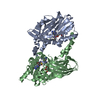
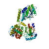
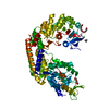
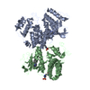
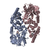
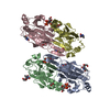
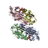
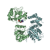
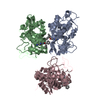
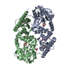
| Title | Crystal structure of human protein arginine methyltransferase PRMT6 bound to SAH and EPZ020411 | ||||||
 Components Components | Protein arginine N-methyltransferase 6 | ||||||
 Keywords Keywords |  TRANSFERASE TRANSFERASE | ||||||
| Function / homology |  Function and homology information Function and homology informationhistone H2AR3 methyltransferase activity / peptidyl-arginine methylation, to asymmetrical-dimethyl arginine / protein-arginine omega-N monomethyltransferase activity / histone H4R3 methyltransferase activity / histone H3R2 methyltransferase activity / type I protein arginine methyltransferase / protein-arginine omega-N asymmetric methyltransferase activity / regulation of megakaryocyte differentiation / histone arginine N-methyltransferase activity / protein-arginine N-methyltransferase activity ...histone H2AR3 methyltransferase activity / peptidyl-arginine methylation, to asymmetrical-dimethyl arginine / protein-arginine omega-N monomethyltransferase activity / histone H4R3 methyltransferase activity / histone H3R2 methyltransferase activity / type I protein arginine methyltransferase / protein-arginine omega-N asymmetric methyltransferase activity / regulation of megakaryocyte differentiation / histone arginine N-methyltransferase activity / protein-arginine N-methyltransferase activity / regulation of mitochondrion organization / histone H3 methyltransferase activity / histone methyltransferase activity / negative regulation of ubiquitin-dependent protein catabolic process / regulation of signal transduction by p53 class mediator / base-excision repair / protein modification process / RUNX1 regulates genes involved in megakaryocyte differentiation and platelet function / RMTs methylate histone arginines / cellular senescence / histone binding / negative regulation of DNA-templated transcription / chromatin binding / nucleolus / negative regulation of transcription by RNA polymerase II / nucleoplasm / nucleusSimilarity search - Function | ||||||
| Biological species |  Homo sapiens (human) Homo sapiens (human) | ||||||
| Method | X-RAY DIFFRACTION / SYNCHROTRON / Resolution: 2.1 Å | ||||||
 Authors Authors | Swinger, K.K. / Boriack-Sjodin, P.A. | ||||||
 Citation Citation | Journal: Acs Med.Chem.Lett. / Year: 2015 Title: Aryl Pyrazoles as Potent Inhibitors of Arginine Methyltransferases: Identification of the First PRMT6 Tool Compound. Authors: Mitchell, L.H. / Drew, A.E. / Ribich, S.A. / Rioux, N. / Swinger, K.K. / Jacques, S.L. / Lingaraj, T. / Boriack-Sjodin, P.A. / Waters, N.J. / Wigle, T.J. / Moradei, O. / Jin, L. / Riera, T. ...Authors: Mitchell, L.H. / Drew, A.E. / Ribich, S.A. / Rioux, N. / Swinger, K.K. / Jacques, S.L. / Lingaraj, T. / Boriack-Sjodin, P.A. / Waters, N.J. / Wigle, T.J. / Moradei, O. / Jin, L. / Riera, T. / Porter-Scott, M. / Moyer, M.P. / Smith, J.J. / Chesworth, R. / Copeland, R.A. | ||||||
| History |
|
- Structure visualization
| Structure viewer | Molecule: MolmilJmol/JSmol |
|---|
- Downloads & links
-Download
| PDBx/mmCIF format | 4y30.cif.gz | 167.3 KB | Display | PDBx/mmCIF format |
|---|---|---|---|---|
| PDB format | pdb4y30.ent.gz | 130.2 KB | Display | PDB format |
| PDBx/mmJSON format | 4y30.json.gz | Tree view | PDBx/mmJSON format | |
| Others |  Other downloads Other downloads |
-Validation report
| Arichive directory | https://data.pdbj.org/pub/pdb/validation_reports/y3/4y30ftp://data.pdbj.org/pub/pdb/validation_reports/y3/4y30 | HTTPS FTP |
|---|
-Related structure data

-Links
 PDBj
PDBj- Assembly
| Deposited unit | 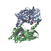
| ||||||||||||||||||
|---|---|---|---|---|---|---|---|---|---|---|---|---|---|---|---|---|---|---|---|
| 1 |
| ||||||||||||||||||
| Unit cell |
| ||||||||||||||||||
| Noncrystallographic symmetry (NCS) | NCS domain:
NCS domain segments: Component-ID: 0 / Ens-ID: 1 / Beg auth comp-ID: THR / Beg label comp-ID: THR / End auth comp-ID: GLU / End label comp-ID: GLU / Refine code: 0 / Auth seq-ID: 39 - 374 / Label seq-ID: 15 - 350
|
-Components
-Protein , 1 types, 2 molecules AB
| #1: Protein | Mass: 39524.836 Da / Num. of mol.: 2 / Fragment: UNP RESIDUES 25-375 Source method: isolated from a genetically manipulated source Source: (gene. exp.) Homo sapiens (human) / Gene: PRMT6, HRMT1L6 / Production host:  Escherichia coli (E. coli) Escherichia coli (E. coli)References: UniProt: Q96LA8, Transferases; Transferring one-carbon groups; Methyltransferases, EC: 2.1.1.125 |
|---|
-Non-polymers , 5 types, 522 molecules 


| #2: Chemical | S-Adenosyl-L-homocysteine Type: L-peptide linking / Mass: 384.411 Da / Num. of mol.: 2 / Source method: obtained synthetically / Formula: C14H20N6O5S Type: L-peptide linking / Mass: 384.411 Da / Num. of mol.: 2 / Source method: obtained synthetically / Formula: C14H20N6O5S#3: Chemical |  Mass: 442.594 Da / Num. of mol.: 2 / Source method: obtained synthetically / Formula: C25H38N4O3 Mass: 442.594 Da / Num. of mol.: 2 / Source method: obtained synthetically / Formula: C25H38N4O3#4: Chemical | ChemComp-MG / |  Mass: 24.305 Da / Num. of mol.: 1 / Source method: obtained synthetically / Formula: Mg Mass: 24.305 Da / Num. of mol.: 1 / Source method: obtained synthetically / Formula: Mg#5: Chemical | Glycerol Mass: 92.094 Da / Num. of mol.: 3 / Source method: obtained synthetically / Formula: C3H8O3 Mass: 92.094 Da / Num. of mol.: 3 / Source method: obtained synthetically / Formula: C3H8O3#6: Water | ChemComp-HOH / | WaterMass: 18.015 Da / Num. of mol.: 514 / Source method: isolated from a natural source / Formula: H2O |
|---|
-Experimental details
-Experiment
| Experiment | Method: X-RAY DIFFRACTION / Number of used crystals: 1 |
|---|
- Sample preparation
| Crystal | Density Matthews: 2.8 Å3/Da / Density % sol: 56.14 % |
|---|---|
| Crystal grow | Temperature: 291.15 K / Method: vapor diffusion, hanging drop / Details: MgCl, MES buffer pH 6.5, Isopropanol, PEG 4000 |
-Data collection
| Diffraction | Mean temperature: 100 K |
|---|---|
| Diffraction source | Source: SYNCHROTRON / Site: SSRF  / Beamline: BL17U / Wavelength: 0.987 Å / Beamline: BL17U / Wavelength: 0.987 Å |
| Detector | Type: ADSC QUANTUM 315r / Detector: CCD / Date: Sep 5, 2014 |
| Radiation | Protocol: SINGLE WAVELENGTH / Monochromatic (M) / Laue (L): M / Scattering type: x-ray |
| Radiation wavelength | Wavelength: 0.987 Å / Relative weight: 1 |
| Reflection | Resolution: 2.1→99.67 Å / Num. obs: 50960 / % possible obs: 99.8 % / Redundancy: 5 % / Net I/σ(I): 8.3 |
- Processing
| Software |
| |||||||||||||||||||||||||||||||||||||||||||||||||||||||||||||||||||||||||||
|---|---|---|---|---|---|---|---|---|---|---|---|---|---|---|---|---|---|---|---|---|---|---|---|---|---|---|---|---|---|---|---|---|---|---|---|---|---|---|---|---|---|---|---|---|---|---|---|---|---|---|---|---|---|---|---|---|---|---|---|---|---|---|---|---|---|---|---|---|---|---|---|---|---|---|---|---|
| Refinement | Resolution: 2.1→43.51 Å / Cor.coef. Fo:Fc: 0.94 / Cor.coef. Fo:Fc free: 0.906 / SU B: 4.729 / SU ML: 0.125 / Cross valid method: THROUGHOUT / σ(F): 0 / ESU R: 0.196 / ESU R Free: 0.169 / Stereochemistry target values: MAXIMUM LIKELIHOOD Details: HYDROGENS HAVE BEEN ADDED IN THE RIDING POSITIONS U VALUES : REFINED INDIVIDUALLY
| |||||||||||||||||||||||||||||||||||||||||||||||||||||||||||||||||||||||||||
| Solvent computation | Ion probe radii: 0.8 Å / Shrinkage radii: 0.8 Å / VDW probe radii: 1.2 Å / Solvent model: MASK | |||||||||||||||||||||||||||||||||||||||||||||||||||||||||||||||||||||||||||
| Displacement parameters | Biso max: 86.74 Å2 / Biso mean: 17.88 Å2 / Biso min: 4.78 Å2
| |||||||||||||||||||||||||||||||||||||||||||||||||||||||||||||||||||||||||||
| Refinement step | Cycle: final / Resolution: 2.1→43.51 Å
| |||||||||||||||||||||||||||||||||||||||||||||||||||||||||||||||||||||||||||
| Refine LS restraints |
| |||||||||||||||||||||||||||||||||||||||||||||||||||||||||||||||||||||||||||
| Refine LS restraints NCS | Ens-ID: 1 / Number: 19680 / Refine-ID: X-RAY DIFFRACTION / Type: interatomic distance / Rms dev position: 0.13 Å / Weight position: 0.05
| |||||||||||||||||||||||||||||||||||||||||||||||||||||||||||||||||||||||||||
| LS refinement shell | Resolution: 2.1→2.154 Å / Total num. of bins used: 20
|
