 Movie
Movie Controller


+ Open data

- Basic information
| Entry | Database: PDB / ID: 1gmy | ||||||
|---|---|---|---|---|---|---|---|
| Title | Cathepsin B complexed with dipeptidyl nitrile inhibitor | ||||||
 Components Components | CATHEPSIN B Cathepsin Cathepsin | ||||||
 Keywords Keywords | HYDROLASE/INHIBITOR / COMPLEX (HYDROLASE-INHIBITOR) / COVALENT COMPLEX / PROTEASE / CATHEPSIN B / HYDROLASE / THIOL PROTEASE / HYDROLASE-INHIBITOR complex | ||||||
| Function / homology |  Function and homology informationcathepsin B / peptidase inhibitor complex / thyroid hormone generation / endolysosome lumen / cellular response to thyroid hormone stimulus / Trafficking and processing of endosomal TLR / proteoglycan binding / Assembly of collagen fibrils and other multimeric structures / Collagen degradation / decidualization ...cathepsin B / peptidase inhibitor complex / thyroid hormone generation / endolysosome lumen / cellular response to thyroid hormone stimulus / Trafficking and processing of endosomal TLR / proteoglycan binding / Assembly of collagen fibrils and other multimeric structures / Collagen degradation / decidualization / collagen catabolic process / cysteine-type peptidase activity / epithelial cell differentiation / collagen binding / MHC class II antigen presentation / proteolysis involved in protein catabolic process / melanosome / peptidase activity / regulation of apoptotic process / collagen-containing extracellular matrix / ficolin-1-rich granule lumen / lysosome / symbiont entry into host cell / apical plasma membrane / external side of plasma membrane / cysteine-type endopeptidase activity / Neutrophil degranulation / perinuclear region of cytoplasm / proteolysis / extracellular space / extracellular exosome / extracellular region Function and homology informationcathepsin B / peptidase inhibitor complex / thyroid hormone generation / endolysosome lumen / cellular response to thyroid hormone stimulus / Trafficking and processing of endosomal TLR / proteoglycan binding / Assembly of collagen fibrils and other multimeric structures / Collagen degradation / decidualization ...cathepsin B / peptidase inhibitor complex / thyroid hormone generation / endolysosome lumen / cellular response to thyroid hormone stimulus / Trafficking and processing of endosomal TLR / proteoglycan binding / Assembly of collagen fibrils and other multimeric structures / Collagen degradation / decidualization / collagen catabolic process / cysteine-type peptidase activity / epithelial cell differentiation / collagen binding / MHC class II antigen presentation / proteolysis involved in protein catabolic process / melanosome / peptidase activity / regulation of apoptotic process / collagen-containing extracellular matrix / ficolin-1-rich granule lumen / lysosome / symbiont entry into host cell / apical plasma membrane / external side of plasma membrane / cysteine-type endopeptidase activity / Neutrophil degranulation / perinuclear region of cytoplasm / proteolysis / extracellular space / extracellular exosome / extracellular regionSimilarity search - Function | ||||||
| Biological species |  HOMO SAPIENS (human) HOMO SAPIENS (human) | ||||||
| Method | X-RAY DIFFRACTION / MOLECULAR REPLACEMENT / Resolution: 1.9 Å | ||||||
 Authors Authors | Greenspan, P.D. / Clark, K.L. / Tommasi, R.A. / Cowen, S.D. / McQuire, L.W. / Farley, D.L. / van Duzer, J.H. / Goldberg, R.L. / Zhou, H. / Du, Z. ...Greenspan, P.D. / Clark, K.L. / Tommasi, R.A. / Cowen, S.D. / McQuire, L.W. / Farley, D.L. / van Duzer, J.H. / Goldberg, R.L. / Zhou, H. / Du, Z. / Fitt, J.J. / Coppa, D.E. / Fang, Z. / Macchia, W. / Zhu, L. / Capparelli, M.P. / Goldstein, R. / Wigg, A.M. / Doughty, J.R. / Bohacek, R.S. / Knap, A.K. | ||||||
 Citation Citation | Journal: J.Med.Chem. / Year: 2001 Title: Identification of Dipeptidyl Nitriles as Potent and Selective Inhibitors of Cathepsin B Through Structure-Based Drug Design Authors: Greenspan, P.D. / Clark, K.L. / Tommasi, R.A. / Cowen, S.D. / Mcquire, L.W. / Farley, D.L. / Van Duzer, J.H. / Goldberg, R.L. / Zhou, H. / Du, Z. / Fitt, J.J. / Coppa, D.E. / Fang, Z. / ...Authors: Greenspan, P.D. / Clark, K.L. / Tommasi, R.A. / Cowen, S.D. / Mcquire, L.W. / Farley, D.L. / Van Duzer, J.H. / Goldberg, R.L. / Zhou, H. / Du, Z. / Fitt, J.J. / Coppa, D.E. / Fang, Z. / Macchia, W. / Zhu, L. / Capparelli, M.P. / Goldstein, R. / Wigg, A.M. / Doughty, J.R. / S Bohacek, R. / Knap, A.K. | ||||||
| History |
| ||||||
| Remark 700 | SHEET THE SHEET STRUCTURE OF THIS MOLECULE IS BIFURCATED. IN ORDER TO REPRESENT THIS FEATURE IN ... SHEET THE SHEET STRUCTURE OF THIS MOLECULE IS BIFURCATED. IN ORDER TO REPRESENT THIS FEATURE IN THE SHEET RECORDS BELOW, TWO SHEETS ARE DEFINED. |
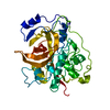
- Structure visualization
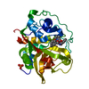
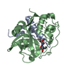
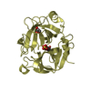
| Structure viewer | Molecule: MolmilJmol/JSmol |
|---|
- Downloads & links
-Download
| PDBx/mmCIF format | 1gmy.cif.gz | 164 KB | Display | PDBx/mmCIF format |
|---|---|---|---|---|
| PDB format | pdb1gmy.ent.gz | 138.5 KB | Display | PDB format |
| PDBx/mmJSON format | 1gmy.json.gz | Tree view | PDBx/mmJSON format | |
| Others |  Other downloads Other downloads |
-Validation report
| Arichive directory | https://data.pdbj.org/pub/pdb/validation_reports/gm/1gmyftp://data.pdbj.org/pub/pdb/validation_reports/gm/1gmy | HTTPS FTP |
|---|
-Related structure data
| Related structure data | |
|---|---|
| Similar structure data |


-Links
 PDBj
PDBj


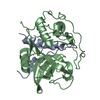
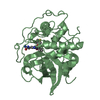
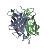
- Assembly
| Deposited unit | 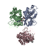
| |||||||||
|---|---|---|---|---|---|---|---|---|---|---|
| 1 | 
| |||||||||
| 2 | 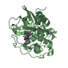
| |||||||||
| 3 | 
| |||||||||
| Unit cell |
| |||||||||
| Components on special symmetry positions |
|
-Components
| #1: Protein | Cathepsin Mass: 28821.123 Da / Num. of mol.: 3 / Fragment: PROTEASE DOMAIN, RESIDUES 80-333 Source method: isolated from a genetically manipulated source Source: (gene. exp.) HOMO SAPIENS (human) / Production host:   SPODOPTERA FRUGIPERDA (fall armyworm) / References: UniProt: P07858, cathepsin B SPODOPTERA FRUGIPERDA (fall armyworm) / References: UniProt: P07858, cathepsin B#2: Chemical |   Mass: 58.082 Da / Num. of mol.: 3 / Source method: obtained synthetically / Formula: C2H6N2 Mass: 58.082 Da / Num. of mol.: 3 / Source method: obtained synthetically / Formula: C2H6N2#3: Chemical |   Mass: 179.216 Da / Num. of mol.: 3 / Source method: obtained synthetically / Formula: C10H13NO2 Mass: 179.216 Da / Num. of mol.: 3 / Source method: obtained synthetically / Formula: C10H13NO2#4: Chemical |   Mass: 212.244 Da / Num. of mol.: 3 / Source method: obtained synthetically / Formula: C14H12O2 Mass: 212.244 Da / Num. of mol.: 3 / Source method: obtained synthetically / Formula: C14H12O2#5: Water | ChemComp-HOH / | Water Mass: 18.015 Da / Num. of mol.: 630 / Source method: isolated from a natural source / Formula: H2O Mass: 18.015 Da / Num. of mol.: 630 / Source method: isolated from a natural source / Formula: H2O |
|---|
-Experimental details
-Experiment
| Experiment | Method: X-RAY DIFFRACTION / Number of used crystals: 1 |
|---|
- Sample preparation
| Crystal | Density Matthews: 2.26 Å3/Da / Density % sol: 45.66 % | |||||||||||||||||||||||||||||||||||
|---|---|---|---|---|---|---|---|---|---|---|---|---|---|---|---|---|---|---|---|---|---|---|---|---|---|---|---|---|---|---|---|---|---|---|---|---|
| Crystal grow | pH: 5.5 / Details: pH 5.50 | |||||||||||||||||||||||||||||||||||
| Crystal grow | *PLUS Temperature: 4 ℃ / Method: vapor diffusion, hanging drop | |||||||||||||||||||||||||||||||||||
| Components of the solutions | *PLUS
|
-Data collection
| Diffraction | Mean temperature: 100 K |
|---|---|
| Diffraction source | Source: ROTATING ANODE / Type: RIGAKU RUH2R / Wavelength: 1.5418 |
| Detector | Type: RIGAKU IMAGE PLATE / Detector: IMAGE PLATE / Date: Apr 15, 1995 / Details: YALE MIRROR |
| Radiation | Monochromator: NI FILTER / Protocol: SINGLE WAVELENGTH / Monochromatic (M) / Laue (L): M / Scattering type: x-ray |
| Radiation wavelength | Wavelength: 1.5418 Å / Relative weight: 1 |
| Reflection | Resolution: 1.9→18 Å / Num. obs: 62966 / % possible obs: 99.9 % / Observed criterion σ(I): -3 / Redundancy: 7.3 % / Biso Wilson estimate: 15.3 Å2 / Rmerge(I) obs: 0.056 / Net I/σ(I): 12.2 |
| Reflection shell | Resolution: 1.9→2.02 Å / Redundancy: 6.5 % / Rmerge(I) obs: 0.305 / Mean I/σ(I) obs: 3.7 / % possible all: 99.8 |
| Reflection | *PLUS Num. obs: 62967 |
| Reflection shell | *PLUS Num. unique obs: 10318 |
- Processing
| Software |
| ||||||||||||||||||||||||||||||||||||||||||||||||||||||||||||||||||||||||||||||||
|---|---|---|---|---|---|---|---|---|---|---|---|---|---|---|---|---|---|---|---|---|---|---|---|---|---|---|---|---|---|---|---|---|---|---|---|---|---|---|---|---|---|---|---|---|---|---|---|---|---|---|---|---|---|---|---|---|---|---|---|---|---|---|---|---|---|---|---|---|---|---|---|---|---|---|---|---|---|---|---|---|---|
| Refinement | Method to determine structure: MOLECULAR REPLACEMENT / Resolution: 1.9→18 Å / Rfactor Rfree error: 0.003 / Isotropic thermal model: BULK SOLVENT MODELING / Cross valid method: THROUGHOUT / σ(F): 1
| ||||||||||||||||||||||||||||||||||||||||||||||||||||||||||||||||||||||||||||||||
| Solvent computation | Solvent model: FLAT MODEL / Bsol: 50.6375 Å2 / ksol: 0.391076 e/Å3 | ||||||||||||||||||||||||||||||||||||||||||||||||||||||||||||||||||||||||||||||||
| Displacement parameters | Biso mean: 22.5 Å2
| ||||||||||||||||||||||||||||||||||||||||||||||||||||||||||||||||||||||||||||||||
| Refine analyze |
| ||||||||||||||||||||||||||||||||||||||||||||||||||||||||||||||||||||||||||||||||
| Refinement step | Cycle: LAST / Resolution: 1.9→18 Å
| ||||||||||||||||||||||||||||||||||||||||||||||||||||||||||||||||||||||||||||||||
| Refine LS restraints |
| ||||||||||||||||||||||||||||||||||||||||||||||||||||||||||||||||||||||||||||||||
| LS refinement shell | Resolution: 1.9→2.02 Å / Rfactor Rfree error: 0.007 / Total num. of bins used: 6
| ||||||||||||||||||||||||||||||||||||||||||||||||||||||||||||||||||||||||||||||||
| Xplor file |
| ||||||||||||||||||||||||||||||||||||||||||||||||||||||||||||||||||||||||||||||||
| Refinement | *PLUS Highest resolution: 1.9 Å / % reflection Rfree: 10 % / Rfactor Rfree: 0.198 / Rfactor Rwork: 0.159 | ||||||||||||||||||||||||||||||||||||||||||||||||||||||||||||||||||||||||||||||||
| Solvent computation | *PLUS | ||||||||||||||||||||||||||||||||||||||||||||||||||||||||||||||||||||||||||||||||
| Displacement parameters | *PLUS | ||||||||||||||||||||||||||||||||||||||||||||||||||||||||||||||||||||||||||||||||
| Refine LS restraints | *PLUS
| ||||||||||||||||||||||||||||||||||||||||||||||||||||||||||||||||||||||||||||||||
| LS refinement shell | *PLUS Rfactor Rfree: 0.206 / Rfactor Rwork: 0.165 |
