 Movie
Movie Controller

[English] 日本語
 Yorodumi
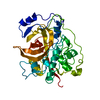
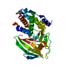
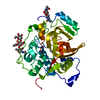
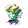
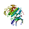
- PDB-2pbh: CRYSTAL STRUCTURE OF HUMAN PROCATHEPSIN B AT 3.3 ANGSTROM RESOLUTION -
+ Open data

- Basic information
| Entry | Database: PDB / ID: 2pbh | ||||||
|---|---|---|---|---|---|---|---|
| Title | CRYSTAL STRUCTURE OF HUMAN PROCATHEPSIN B AT 3.3 ANGSTROM RESOLUTION | ||||||
 Components Components | PROCATHEPSIN B | ||||||
 Keywords Keywords |  THIOL PROTEASE / CATHEPSIN B / CYSTEINE PROTEASE / PROENZYME / PAPAIN THIOL PROTEASE / CATHEPSIN B / CYSTEINE PROTEASE / PROENZYME / PAPAIN | ||||||
| Function / homology |  Function and homology informationcathepsin B / peptidase inhibitor complex / thyroid hormone generation / endolysosome lumen / cellular response to thyroid hormone stimulus / Trafficking and processing of endosomal TLR / proteoglycan binding / Assembly of collagen fibrils and other multimeric structures / Collagen degradation / decidualization ...cathepsin B / peptidase inhibitor complex / thyroid hormone generation / endolysosome lumen / cellular response to thyroid hormone stimulus / Trafficking and processing of endosomal TLR / proteoglycan binding / Assembly of collagen fibrils and other multimeric structures / Collagen degradation / decidualization / collagen catabolic process / cysteine-type peptidase activity / epithelial cell differentiation / collagen binding / MHC class II antigen presentation / proteolysis involved in protein catabolic process / melanosome / peptidase activity / regulation of apoptotic process / collagen-containing extracellular matrix / ficolin-1-rich granule lumen / lysosome / symbiont entry into host cell / apical plasma membrane / external side of plasma membrane / cysteine-type endopeptidase activity / Neutrophil degranulation / perinuclear region of cytoplasm / proteolysis / extracellular space / extracellular exosome / extracellular region Function and homology informationcathepsin B / peptidase inhibitor complex / thyroid hormone generation / endolysosome lumen / cellular response to thyroid hormone stimulus / Trafficking and processing of endosomal TLR / proteoglycan binding / Assembly of collagen fibrils and other multimeric structures / Collagen degradation / decidualization ...cathepsin B / peptidase inhibitor complex / thyroid hormone generation / endolysosome lumen / cellular response to thyroid hormone stimulus / Trafficking and processing of endosomal TLR / proteoglycan binding / Assembly of collagen fibrils and other multimeric structures / Collagen degradation / decidualization / collagen catabolic process / cysteine-type peptidase activity / epithelial cell differentiation / collagen binding / MHC class II antigen presentation / proteolysis involved in protein catabolic process / melanosome / peptidase activity / regulation of apoptotic process / collagen-containing extracellular matrix / ficolin-1-rich granule lumen / lysosome / symbiont entry into host cell / apical plasma membrane / external side of plasma membrane / cysteine-type endopeptidase activity / Neutrophil degranulation / perinuclear region of cytoplasm / proteolysis / extracellular space / extracellular exosome / extracellular regionSimilarity search - Function | ||||||
| Biological species |  Homo sapiens (human) Homo sapiens (human) | ||||||
| Method | X-RAY DIFFRACTION / MOLECULAR REPLACEMENT / Resolution: 3.3 Å | ||||||
 Authors Authors | Podobnik, M. / Turk, D. / Kuhelj, R. / Turk, V. | ||||||
 Citation Citation | Journal: FEBS Lett. / Year: 1996 Title: Crystal structures of human procathepsin B at 3.2 and 3.3 Angstroms resolution reveal an interaction motif between a papain-like cysteine protease and its propeptide. Authors: Turk, D. / Podobnik, M. / Kuhelj, R. / Dolinar, M. / Turk, V. | ||||||
| History |
|
- Structure visualization
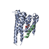
| Structure viewer | Molecule: MolmilJmol/JSmol |
|---|
- Downloads & links
-Download
| PDBx/mmCIF format | 2pbh.cif.gz | 77.6 KB | Display | PDBx/mmCIF format |
|---|---|---|---|---|
| PDB format | pdb2pbh.ent.gz | 57.2 KB | Display | PDB format |
| PDBx/mmJSON format | 2pbh.json.gz | Tree view | PDBx/mmJSON format | |
| Others |  Other downloads Other downloads |
-Validation report
| Arichive directory | https://data.pdbj.org/pub/pdb/validation_reports/pb/2pbhftp://data.pdbj.org/pub/pdb/validation_reports/pb/2pbh | HTTPS FTP |
|---|
-Related structure data


-Links
 PDBj
PDBj


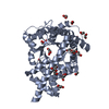
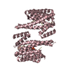
- Assembly
| Deposited unit | 
| ||||||||
|---|---|---|---|---|---|---|---|---|---|
| 1 |
| ||||||||
| Unit cell |
|
-Components
| #1: Protein | Mass: 35226.391 Da / Num. of mol.: 1 Source method: isolated from a genetically manipulated source Source: (gene. exp.) Homo sapiens (human) / Cell line: BL21 / Plasmid: PET3A / Production host:  Escherichia coli (E. coli) / Strain (production host): BL21 (DE3) PLYSS / References: UniProt: P07858, cathepsin B Escherichia coli (E. coli) / Strain (production host): BL21 (DE3) PLYSS / References: UniProt: P07858, cathepsin B |
|---|
-Experimental details
-Experiment
| Experiment | Method: X-RAY DIFFRACTION / Number of used crystals: 2 |
|---|
- Sample preparation
| Crystal | Density Matthews: 2.35 Å3/Da / Density % sol: 48 % | |||||||||||||||
|---|---|---|---|---|---|---|---|---|---|---|---|---|---|---|---|---|
| Crystal grow | pH: 7.2 Details: PROTEIN WAS CRYSTALLIZED FROM 2 M AMMONIUM SULFATE, 700 MM TRIS, PH 7.2 | |||||||||||||||
| Crystal grow | *PLUS Temperature: 20 ℃ / pH: 7.1 / Method: vapor diffusion, sitting drop | |||||||||||||||
| Components of the solutions | *PLUS
|
-Data collection
| Diffraction | Mean temperature: 283 K |
|---|---|
| Diffraction source | Source: ROTATING ANODE / Type: RIGAKU RUH2R / Wavelength: 1.5418 |
| Detector | Type: MARRESEARCH / Detector: IMAGE PLATE / Date: Oct 1, 1995 / Details: COLLIMATOR |
| Radiation | Monochromator: NI FILTER / Monochromatic (M) / Laue (L): M / Scattering type: x-ray |
| Radiation wavelength | Wavelength: 1.5418 Å / Relative weight: 1 |
| Reflection | Resolution: 3.3→8 Å / Num. obs: 4297 / % possible obs: 94.2 % / Observed criterion σ(I): 2 / Redundancy: 8.5 % / Rmerge(I) obs: 0.186 / Rsym value: 0.08 / Net I/σ(I): 3.9 |
| Reflection shell | Resolution: 3.3→3.4 Å / Redundancy: 4.2 % / Rmerge(I) obs: 0.066 / Mean I/σ(I) obs: 2.4 / Rsym value: 0.316 / % possible all: 96.7 |
- Processing
| Software |
| ||||||||||||||||
|---|---|---|---|---|---|---|---|---|---|---|---|---|---|---|---|---|---|
| Refinement | Method to determine structure: MOLECULAR REPLACEMENT Starting model: PDB ENTRY Resolution: 3.3→10 Å / σ(F): 2
| ||||||||||||||||
| Refinement step | Cycle: LAST / Resolution: 3.3→10 Å
| ||||||||||||||||
| Software | *PLUS Name: MAIN / Classification: refinement | ||||||||||||||||
| Refinement | *PLUS Num. reflection all: 4297 / Rfactor Rfree: 0.269 | ||||||||||||||||
| Solvent computation | *PLUS | ||||||||||||||||
| Displacement parameters | *PLUS | ||||||||||||||||
| Refine LS restraints | *PLUS
|
