 Movie
Movie Controller


+ Open data

- Basic information
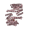
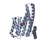
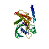
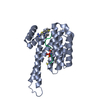
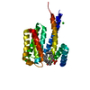
| Entry | Database: PDB / ID: 5f74 | ||||||
|---|---|---|---|---|---|---|---|
| Title | Crystal structure of ChREBP:14-3-3 complex bound with AMP | ||||||
 Components Components |
| ||||||
 Keywords Keywords |  TRANSCRIPTION / ChREBP / 14-3-3 / AMP / Allosteric TRANSCRIPTION / ChREBP / 14-3-3 / AMP / Allosteric | ||||||
| Function / homology |  Function and homology information Function and homology informationPKA-mediated phosphorylation of key metabolic factors / : / Butyrate Response Factor 1 (BRF1) binds and destabilizes mRNA / carbohydrate response element binding / Tristetraprolin (TTP, ZFP36) binds and destabilizes mRNA / Regulation of localization of FOXO transcription factors / Chk1/Chk2(Cds1) mediated inactivation of Cyclin B:Cdk1 complex / RHO GTPases activate PKNs / regulation of cell cycle => GO:0051726 / Activation of BAD and translocation to mitochondria ...PKA-mediated phosphorylation of key metabolic factors / : / Butyrate Response Factor 1 (BRF1) binds and destabilizes mRNA / carbohydrate response element binding / Tristetraprolin (TTP, ZFP36) binds and destabilizes mRNA / Regulation of localization of FOXO transcription factors / Chk1/Chk2(Cds1) mediated inactivation of Cyclin B:Cdk1 complex / RHO GTPases activate PKNs / regulation of cell cycle => GO:0051726 / Activation of BAD and translocation to mitochondria / Frs2-mediated activation / MTOR signalling / Signaling by Hippo / Negative regulation of MAPK pathway / negative regulation of protein dephosphorylation / glucose mediated signaling pathway / TP53 Regulates Metabolic Genes / RAF activation / mTORC1-mediated signalling / MAP2K and MAPK activation / Rap1 signalling / cytoplasmic sequestering of protein / negative regulation of G protein-coupled receptor signaling pathway / positive regulation of fatty acid biosynthetic process / negative regulation of oxidative phosphorylation / regulation of glycolytic process / protein kinase inhibitor activity / transcription factor binding / phosphoserine residue binding / protein targeting / response to glucose / fatty acid homeostasis / energy homeostasis / negative regulation of peptidyl-serine phosphorylation / positive regulation of lipid biosynthetic process / transcription repressor complex / positive regulation of glycolytic process / cellular response to glucose stimulus / phosphoprotein binding / intracellular protein transport / DNA-binding transcription repressor activity, RNA polymerase II-specific / histone deacetylase binding / melanosome / glucose homeostasis / transcription regulator complex / sequence-specific DNA binding / DNA-binding transcription factor activity, RNA polymerase II-specific / RNA polymerase II cis-regulatory region sequence-specific DNA binding / DNA-binding transcription factor activity / protein heterodimerization activity / protein domain specific binding / negative regulation of DNA-templated transcription / positive regulation of cell population proliferation / protein-containing complex binding / regulation of transcription by RNA polymerase II / protein kinase binding / perinuclear region of cytoplasm / positive regulation of DNA-templated transcription / negative regulation of transcription by RNA polymerase II / enzyme binding / signal transduction / positive regulation of transcription by RNA polymerase II / protein-containing complex / DNA binding / nucleoplasm / identical protein binding / nucleus / cytosol / cytoplasmSimilarity search - Function | ||||||
| Biological species |  Mus musculus (house mouse)Rattus norvegicus (Norway rat) Mus musculus (house mouse)Rattus norvegicus (Norway rat) | ||||||
| Method | X-RAY DIFFRACTION / SYNCHROTRON / MOLECULAR REPLACEMENT / Resolution: 2.35 Å | ||||||
 Authors Authors | Jung, H. / Uyeda, K. | ||||||
 Citation Citation | Journal: J.Biol.Chem. / Year: 2016 Title: Metabolite Regulation of Nuclear Localization of Carbohydrate-response Element-binding Protein (ChREBP): ROLE OF AMP AS AN ALLOSTERIC INHIBITOR. Authors: Sato, S. / Jung, H. / Nakagawa, T. / Pawlosky, R. / Takeshima, T. / Lee, W.R. / Sakiyama, H. / Laxman, S. / Wynn, R.M. / Tu, B.P. / MacMillan, J.B. / De Brabander, J.K. / Veech, R.L. / Uyeda, K. | ||||||
| History |
|
- Structure visualization
| Structure viewer | Molecule: MolmilJmol/JSmol |
|---|
- Downloads & links
-Download
| PDBx/mmCIF format | 5f74.cif.gz | 69.6 KB | Display | PDBx/mmCIF format |
|---|---|---|---|---|
| PDB format | pdb5f74.ent.gz | 48.9 KB | Display | PDB format |
| PDBx/mmJSON format | 5f74.json.gz | Tree view | PDBx/mmJSON format | |
| Others |  Other downloads Other downloads |
-Validation report
| Arichive directory | https://data.pdbj.org/pub/pdb/validation_reports/f7/5f74ftp://data.pdbj.org/pub/pdb/validation_reports/f7/5f74 | HTTPS FTP |
|---|
-Related structure data
| Related structure data | 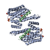 4gntS S: Starting model for refinement |
|---|---|
| Similar structure data |


-Links
 PDBj
PDBj


- Assembly
| Deposited unit | 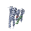
| ||||||||
|---|---|---|---|---|---|---|---|---|---|
| 1 |
| ||||||||
| 2 | 
| ||||||||
| Unit cell |
| ||||||||
| Components on special symmetry positions |
|
-Components
| #1: Protein | Mass: 28118.404 Da / Num. of mol.: 1 Source method: isolated from a genetically manipulated source Source: (gene. exp.) Mus musculus (house mouse) / Gene: Ywhab / Plasmid: pET Duet / Production host:  Escherichia coli (E. coli) / References: UniProt: Q9CQV8 Escherichia coli (E. coli) / References: UniProt: Q9CQV8 |
|---|---|
| #2: Protein | / ChREBP / Class D basic helix-loop-helix protein 14 / bHLHd14 / MLX interactor / MLX-interacting ...ChREBP / Class D basic helix-loop-helix protein 14 / bHLHd14 / MLX interactor / MLX-interacting protein-like / WS basic-helix-loop-helix leucine zipper protein / WS-bHLH / Williams-Beuren syndrome chromosomal region 14 protein Mass: 22656.010 Da / Num. of mol.: 1 / Fragment: UNP residues 1-196 Source method: isolated from a genetically manipulated source Source: (gene. exp.) Rattus norvegicus (Norway rat) / Gene: Mlxipl, Wbscr14 / Plasmid: pET Duet / Production host: Escherichia coli (E. coli) / References: UniProt: Q8VIP2 |
| #3: Chemical | ChemComp-AMP / Adenosine monophosphate  Mass: 347.221 Da / Num. of mol.: 1 / Source method: obtained synthetically / Formula: C10H14N5O7P / Comment: AMP*YM Mass: 347.221 Da / Num. of mol.: 1 / Source method: obtained synthetically / Formula: C10H14N5O7P / Comment: AMP*YM |
| #4: Water | ChemComp-HOH / Water Mass: 18.015 Da / Num. of mol.: 45 / Source method: isolated from a natural source / Formula: H2O Mass: 18.015 Da / Num. of mol.: 45 / Source method: isolated from a natural source / Formula: H2O |
-Experimental details
-Experiment
| Experiment | Method: X-RAY DIFFRACTION |
|---|
- Sample preparation
| Crystal | Density Matthews: 1.95 Å3/Da / Density % sol: 37.05 % |
|---|---|
| Crystal grow | Temperature: 293 K / Method: vapor diffusion, hanging drop Details: 100 mM sodium citrate 200 mM sodium potassium tartrate 2.2 M ammonium sulfate PH range: 6.2 -7.0 |
-Data collection
| Diffraction | Mean temperature: 120 K |
|---|---|
| Diffraction source | Source: SYNCHROTRON / Site: APS  / Beamline: 19-ID / Wavelength: 1 Å / Beamline: 19-ID / Wavelength: 1 Å |
| Detector | Type: ADSC QUANTUM 315 / Detector: CCD / Date: Jul 16, 2015 |
| Radiation | Protocol: SINGLE WAVELENGTH / Monochromatic (M) / Laue (L): M / Scattering type: x-ray |
| Radiation wavelength | Wavelength: 1 Å / Relative weight: 1 |
| Reflection | Resolution: 2.35→50 Å / Num. obs: 17340 / % possible obs: 100 % / Redundancy: 6.5 % / Rmerge(I) obs: 0.041 / Net I/σ(I): 22.9 |
- Processing
| Software |
| |||||||||||||||||||||||||||||||||||||||||||||||||
|---|---|---|---|---|---|---|---|---|---|---|---|---|---|---|---|---|---|---|---|---|---|---|---|---|---|---|---|---|---|---|---|---|---|---|---|---|---|---|---|---|---|---|---|---|---|---|---|---|---|---|
| Refinement | Method to determine structure: MOLECULAR REPLACEMENT Starting model: 4GNT Resolution: 2.35→41.419 Å / SU ML: 0.3 / Cross valid method: FREE R-VALUE / σ(F): 1.33 / Phase error: 25.25 / Stereochemistry target values: ML
| |||||||||||||||||||||||||||||||||||||||||||||||||
| Solvent computation | Shrinkage radii: 0.9 Å / VDW probe radii: 1.11 Å / Solvent model: FLAT BULK SOLVENT MODEL | |||||||||||||||||||||||||||||||||||||||||||||||||
| Refinement step | Cycle: LAST / Resolution: 2.35→41.419 Å
| |||||||||||||||||||||||||||||||||||||||||||||||||
| Refine LS restraints |
| |||||||||||||||||||||||||||||||||||||||||||||||||
| LS refinement shell |
|
