 Movie
Movie Controller

[English] 日本語
 Yorodumi
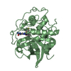
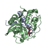
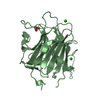
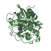
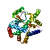
- PDB-1huc: THE REFINED 2.15 ANGSTROMS X-RAY CRYSTAL STRUCTURE OF HUMAN LIVER... -
+ Open data

- Basic information
| Entry | Database: PDB / ID: 1huc | ||||||
|---|---|---|---|---|---|---|---|
| Title | THE REFINED 2.15 ANGSTROMS X-RAY CRYSTAL STRUCTURE OF HUMAN LIVER CATHEPSIN B: THE STRUCTURAL BASIS FOR ITS SPECIFICITY | ||||||
 Components Components | (CATHEPSIN B Cathepsin) x 2 Cathepsin) x 2 | ||||||
 Keywords Keywords | THIOL PROTEASE | ||||||
| Function / homology |  Function and homology informationcathepsin B / peptidase inhibitor complex / thyroid hormone generation / endolysosome lumen / cellular response to thyroid hormone stimulus / Trafficking and processing of endosomal TLR / proteoglycan binding / Assembly of collagen fibrils and other multimeric structures / Collagen degradation / decidualization ...cathepsin B / peptidase inhibitor complex / thyroid hormone generation / endolysosome lumen / cellular response to thyroid hormone stimulus / Trafficking and processing of endosomal TLR / proteoglycan binding / Assembly of collagen fibrils and other multimeric structures / Collagen degradation / decidualization / collagen catabolic process / cysteine-type peptidase activity / epithelial cell differentiation / collagen binding / MHC class II antigen presentation / proteolysis involved in protein catabolic process / melanosome / peptidase activity / regulation of apoptotic process / collagen-containing extracellular matrix / ficolin-1-rich granule lumen / lysosome / symbiont entry into host cell / apical plasma membrane / external side of plasma membrane / cysteine-type endopeptidase activity / Neutrophil degranulation / perinuclear region of cytoplasm / proteolysis / extracellular space / extracellular exosome / extracellular region Function and homology informationcathepsin B / peptidase inhibitor complex / thyroid hormone generation / endolysosome lumen / cellular response to thyroid hormone stimulus / Trafficking and processing of endosomal TLR / proteoglycan binding / Assembly of collagen fibrils and other multimeric structures / Collagen degradation / decidualization ...cathepsin B / peptidase inhibitor complex / thyroid hormone generation / endolysosome lumen / cellular response to thyroid hormone stimulus / Trafficking and processing of endosomal TLR / proteoglycan binding / Assembly of collagen fibrils and other multimeric structures / Collagen degradation / decidualization / collagen catabolic process / cysteine-type peptidase activity / epithelial cell differentiation / collagen binding / MHC class II antigen presentation / proteolysis involved in protein catabolic process / melanosome / peptidase activity / regulation of apoptotic process / collagen-containing extracellular matrix / ficolin-1-rich granule lumen / lysosome / symbiont entry into host cell / apical plasma membrane / external side of plasma membrane / cysteine-type endopeptidase activity / Neutrophil degranulation / perinuclear region of cytoplasm / proteolysis / extracellular space / extracellular exosome / extracellular regionSimilarity search - Function | ||||||
| Biological species |  Homo sapiens (human) Homo sapiens (human) | ||||||
| Method | X-RAY DIFFRACTION / Resolution: 2.1 Å | ||||||
 Authors Authors | Musil, D. / Bode, W. | ||||||
 Citation Citation | Journal: EMBO J. / Year: 1991 Title: The refined 2.15 A X-ray crystal structure of human liver cathepsin B: the structural basis for its specificity. Authors: Musil, D. / Zucic, D. / Turk, D. / Engh, R.A. / Mayr, I. / Huber, R. / Popovic, T. / Turk, V. / Towatari, T. / Katunuma, N. / Bode, W. | ||||||
| History |
|
- Structure visualization
| Structure viewer | Molecule: MolmilJmol/JSmol |
|---|
- Downloads & links
-Download
| PDBx/mmCIF format | 1huc.cif.gz | 112.4 KB | Display | PDBx/mmCIF format |
|---|---|---|---|---|
| PDB format | pdb1huc.ent.gz | 87.4 KB | Display | PDB format |
| PDBx/mmJSON format | 1huc.json.gz | Tree view | PDBx/mmJSON format | |
| Others |  Other downloads Other downloads |
-Validation report
| Arichive directory | https://data.pdbj.org/pub/pdb/validation_reports/hu/1hucftp://data.pdbj.org/pub/pdb/validation_reports/hu/1huc | HTTPS FTP |
|---|
-Related structure data
| Similar structure data |
|---|


-Links
 PDBj
PDBj


- Assembly
| Deposited unit | 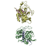
| ||||||||
|---|---|---|---|---|---|---|---|---|---|
| 1 | 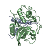
| ||||||||
| 2 | 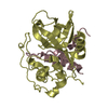
| ||||||||
| Unit cell |
| ||||||||
| Atom site foot note | 1: ASN B 113 - GLY B 114 OMEGA = 210.44 PEPTIDE BOND DEVIATES SIGNIFICANTLY FROM TRANS CONFORMATION 2: CIS PROLINE - PRO B 138 / 3: CIS PROLINE - PRO D 138 | ||||||||
| Noncrystallographic symmetry (NCS) | NCS oper: (Code: given Matrix: (-0.019, -0.0043, -0.9998), Vector : Details | THE TRANSFORMATION PRESENTED ON *MTRIX* RECORDS BELOW WILL YIELD APPROXIMATE COORDINATES FOR CHAINS *A* AND *B* WHEN APPLIED TO CHAINS *C* AND *D*, RESPECTIVELY. | |
-Components
| #1: Protein/peptide | Cathepsin Mass: 5213.840 Da / Num. of mol.: 2 Source method: isolated from a genetically manipulated source Source: (gene. exp.) Homo sapiens (human) / References: UniProt: P07858, cathepsin B#2: Protein | CathepsinMass: 22437.910 Da / Num. of mol.: 2 Source method: isolated from a genetically manipulated source Source: (gene. exp.) Homo sapiens (human) / References: UniProt: P07858, cathepsin B#3: Water | ChemComp-HOH / | Water Mass: 18.015 Da / Num. of mol.: 283 / Source method: isolated from a natural source / Formula: H2O Mass: 18.015 Da / Num. of mol.: 283 / Source method: isolated from a natural source / Formula: H2O |
|---|
-Experimental details
-Experiment
| Experiment | Method: X-RAY DIFFRACTION |
|---|
- Sample preparation
| Crystal | Density Matthews: 2.22 Å3/Da / Density % sol: 44.59 % | |||||||||||||||
|---|---|---|---|---|---|---|---|---|---|---|---|---|---|---|---|---|
| Crystal grow | *PLUS Temperature: 20 ℃ / Method: vapor diffusion, hanging drop | |||||||||||||||
| Components of the solutions | *PLUS
|
-Data collection
| Radiation | Scattering type: x-ray |
|---|---|
| Radiation wavelength | Relative weight: 1 |
- Processing
| Software |
| ||||||||||||
|---|---|---|---|---|---|---|---|---|---|---|---|---|---|
| Refinement | Rfactor obs: 0.164 / Highest resolution: 2.1 Å | ||||||||||||
| Refinement step | Cycle: LAST / Highest resolution: 2.1 Å
| ||||||||||||
| Refinement | *PLUS Highest resolution: 2.15 Å / Lowest resolution: 8 Å / Rfactor obs: 0.164 | ||||||||||||
| Solvent computation | *PLUS | ||||||||||||
| Displacement parameters | *PLUS | ||||||||||||
| Refine LS restraints | *PLUS
|
