 Movie
Movie Controller


+ Open data

- Basic information
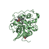
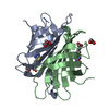
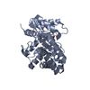
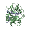
| Entry | Database: PDB / ID: 3ai8 | ||||||
|---|---|---|---|---|---|---|---|
| Title | Cathepsin B in complex with the nitroxoline | ||||||
 Components Components | Cathepsin B | ||||||
 Keywords Keywords | HYDROLASE/HYDROLASE INHIBITOR / cathepsin B / reversible inhibitor / Nitroxoline / 8-Hydroxy-5-nitroquinoline / HYDROLASE-HYDROLASE INHIBITOR complex | ||||||
| Function / homology |  Function and homology informationcathepsin B / peptidase inhibitor complex / thyroid hormone generation / endolysosome lumen / cellular response to thyroid hormone stimulus / Trafficking and processing of endosomal TLR / proteoglycan binding / Assembly of collagen fibrils and other multimeric structures / Collagen degradation / decidualization ...cathepsin B / peptidase inhibitor complex / thyroid hormone generation / endolysosome lumen / cellular response to thyroid hormone stimulus / Trafficking and processing of endosomal TLR / proteoglycan binding / Assembly of collagen fibrils and other multimeric structures / Collagen degradation / decidualization / collagen catabolic process / cysteine-type peptidase activity / epithelial cell differentiation / collagen binding / MHC class II antigen presentation / proteolysis involved in protein catabolic process / melanosome / peptidase activity / regulation of apoptotic process / collagen-containing extracellular matrix / ficolin-1-rich granule lumen / lysosome / symbiont entry into host cell / apical plasma membrane / external side of plasma membrane / cysteine-type endopeptidase activity / Neutrophil degranulation / perinuclear region of cytoplasm / proteolysis / extracellular space / extracellular exosome / extracellular region Function and homology informationcathepsin B / peptidase inhibitor complex / thyroid hormone generation / endolysosome lumen / cellular response to thyroid hormone stimulus / Trafficking and processing of endosomal TLR / proteoglycan binding / Assembly of collagen fibrils and other multimeric structures / Collagen degradation / decidualization ...cathepsin B / peptidase inhibitor complex / thyroid hormone generation / endolysosome lumen / cellular response to thyroid hormone stimulus / Trafficking and processing of endosomal TLR / proteoglycan binding / Assembly of collagen fibrils and other multimeric structures / Collagen degradation / decidualization / collagen catabolic process / cysteine-type peptidase activity / epithelial cell differentiation / collagen binding / MHC class II antigen presentation / proteolysis involved in protein catabolic process / melanosome / peptidase activity / regulation of apoptotic process / collagen-containing extracellular matrix / ficolin-1-rich granule lumen / lysosome / symbiont entry into host cell / apical plasma membrane / external side of plasma membrane / cysteine-type endopeptidase activity / Neutrophil degranulation / perinuclear region of cytoplasm / proteolysis / extracellular space / extracellular exosome / extracellular regionSimilarity search - Function | ||||||
| Biological species |  Homo sapiens (human) Homo sapiens (human) | ||||||
| Method | X-RAY DIFFRACTION / SYNCHROTRON / MOLECULAR REPLACEMENT / Resolution: 2.11 Å | ||||||
 Authors Authors | Renko, M. / Mirkovic, B. / Gobec, S. / Kos, J. / Turk, D. | ||||||
 Citation Citation | Journal: Chemmedchem / Year: 2011 Title: Novel mechanism of cathepsin B inhibition by antibiotic nitroxoline and related compounds Authors: Mirkovic, B. / Renko, M. / Turk, S. / Sosic, I. / Jevnikar, Z. / Obermajer, N. / Turk, D. / Gobec, S. / Kos, J. | ||||||
| History |
|
- Structure visualization
| Structure viewer | Molecule: MolmilJmol/JSmol |
|---|
- Downloads & links
-Download
| PDBx/mmCIF format | 3ai8.cif.gz | 116 KB | Display | PDBx/mmCIF format |
|---|---|---|---|---|
| PDB format | pdb3ai8.ent.gz | 89.4 KB | Display | PDB format |
| PDBx/mmJSON format | 3ai8.json.gz | Tree view | PDBx/mmJSON format | |
| Others |  Other downloads Other downloads |
-Validation report
| Arichive directory | https://data.pdbj.org/pub/pdb/validation_reports/ai/3ai8ftp://data.pdbj.org/pub/pdb/validation_reports/ai/3ai8 | HTTPS FTP |
|---|
-Related structure data
| Related structure data | 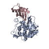 3k9mS S: Starting model for refinement |
|---|---|
| Similar structure data |


-Links
 PDBj
PDBj


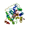
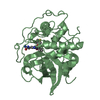
- Assembly
| Deposited unit | 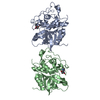
| ||||||||
|---|---|---|---|---|---|---|---|---|---|
| 1 | 
| ||||||||
| 2 | 
| ||||||||
| Unit cell |
|
-Components
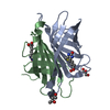
| #1: Protein | / Cathepsin B1 / APP secretase / APPS / Cathepsin B light chain / Cathepsin B heavy chain Mass: 28085.316 Da / Num. of mol.: 2 Source method: isolated from a genetically manipulated source Source: (gene. exp.) Homo sapiens (human) / Gene: CTSB / Plasmid: PET3a / Production host:  Escherichia coli (E. coli) / Strain (production host): Bl21de3 / References: UniProt: P07858, cathepsin B Escherichia coli (E. coli) / Strain (production host): Bl21de3 / References: UniProt: P07858, cathepsin B#2: Chemical | Nitroxoline  Mass: 190.156 Da / Num. of mol.: 2 / Source method: obtained synthetically / Formula: C9H6N2O3 / Comment: antibiotic*YM Mass: 190.156 Da / Num. of mol.: 2 / Source method: obtained synthetically / Formula: C9H6N2O3 / Comment: antibiotic*YM#3: Water | ChemComp-HOH / | Water Mass: 18.015 Da / Num. of mol.: 284 / Source method: isolated from a natural source / Formula: H2O Mass: 18.015 Da / Num. of mol.: 284 / Source method: isolated from a natural source / Formula: H2O |
|---|
-Experimental details
-Experiment
| Experiment | Method: X-RAY DIFFRACTION / Number of used crystals: 1 |
|---|
- Sample preparation
| Crystal | Density Matthews: 1.97 Å3/Da / Density % sol: 37.59 % |
|---|---|
| Crystal grow | Temperature: 293 K / Method: vapor diffusion, sitting drop / pH: 3 Details: 0.1M citric acid, 21% PEG6000, pH 3.0, VAPOR DIFFUSION, SITTING DROP, temperature 293K |
-Data collection
| Diffraction | Mean temperature: 100 K |
|---|---|
| Diffraction source | Source: SYNCHROTRON / Site: ELETTRA  / Beamline: 5.2R / Wavelength: 1 Å / Beamline: 5.2R / Wavelength: 1 Å |
| Detector | Type: PSI PILATUS 6M / Detector: PIXEL / Date: Apr 5, 2010 / Details: Collimating and focusing, Pt-coated mirrors |
| Radiation | Monochromator: Double Crystal Si111 / Protocol: SINGLE WAVELENGTH / Monochromatic (M) / Laue (L): M / Scattering type: x-ray |
| Radiation wavelength | Wavelength: 1 Å / Relative weight: 1 |
| Reflection | Resolution: 2.1→30 Å / Num. all: 25966 / Num. obs: 25966 / % possible obs: 92.5 % / Observed criterion σ(F): 1 / Observed criterion σ(I): 1 / Redundancy: 4.3 % / Rmerge(I) obs: 0.096 |
| Reflection shell | Resolution: 2.1→2.14 Å / Redundancy: 4.1 % / Rmerge(I) obs: 0.31 / Mean I/σ(I) obs: 4.2 / % possible all: 96.2 |
- Processing
| Software |
| |||||||||||||||||||||||||||||||||||||||||||||||||||||||||||||||||
|---|---|---|---|---|---|---|---|---|---|---|---|---|---|---|---|---|---|---|---|---|---|---|---|---|---|---|---|---|---|---|---|---|---|---|---|---|---|---|---|---|---|---|---|---|---|---|---|---|---|---|---|---|---|---|---|---|---|---|---|---|---|---|---|---|---|---|
| Refinement | Method to determine structure: MOLECULAR REPLACEMENT Starting model: 3K9M Resolution: 2.11→29.1 Å / Cor.coef. Fo:Fc: 0.946 / WRfactor Rwork: 0.178 / Occupancy max: 1 / Occupancy min: 0 / FOM work R set: 0.881 / SU R Cruickshank DPI: 0.36 / σ(F): 0 / Stereochemistry target values: MAXIMUM LIKELIHOOD Details: HYDROGENS HAVE BEEN ADDED IN THE RIDING POSITIONS U VALUES: REFINED INDIVIDUALLY. The structure was refined also with MAIN
| |||||||||||||||||||||||||||||||||||||||||||||||||||||||||||||||||
| Solvent computation | Ion probe radii: 0.8 Å / Shrinkage radii: 0.8 Å / VDW probe radii: 1.4 Å / Solvent model: MASK | |||||||||||||||||||||||||||||||||||||||||||||||||||||||||||||||||
| Displacement parameters | Biso max: 76.93 Å2 / Biso mean: 22.877 Å2 / Biso min: 6.05 Å2
| |||||||||||||||||||||||||||||||||||||||||||||||||||||||||||||||||
| Refinement step | Cycle: LAST / Resolution: 2.11→29.1 Å
| |||||||||||||||||||||||||||||||||||||||||||||||||||||||||||||||||
| Refine LS restraints |
| |||||||||||||||||||||||||||||||||||||||||||||||||||||||||||||||||
| LS refinement shell | Resolution: 2.107→2.162 Å / Total num. of bins used: 20
|
