 Movie
Movie Controller


+ Open data

- Basic information
| Entry | Database: PDB / ID: 4xzs | |||||||||
|---|---|---|---|---|---|---|---|---|---|---|
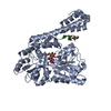
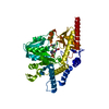
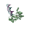
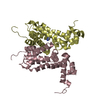
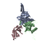
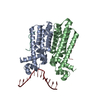
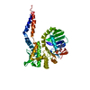
| Title | Crystal Structure of TRIAP1-MBP fusion | |||||||||
 Components Components | Maltose-binding periplasmic protein,TP53-regulated inhibitor of apoptosis 1 | |||||||||
 Keywords Keywords |  APOPTOSIS / lipid / cX9cX motif / cancer / mitochondria / chaperone APOPTOSIS / lipid / cX9cX motif / cancer / mitochondria / chaperone | |||||||||
| Function / homology |  Function and homology information Function and homology informationregulation of membrane lipid distribution / phosphatidic acid transfer activity / positive regulation of phospholipid transport / phospholipid transport / phospholipid translocation / detection of maltose stimulus / negative regulation of intrinsic apoptotic signaling pathway in response to DNA damage by p53 class mediator / maltose transport complex / negative regulation of release of cytochrome c from mitochondria / maltose binding ...regulation of membrane lipid distribution / phosphatidic acid transfer activity / positive regulation of phospholipid transport / phospholipid transport / phospholipid translocation / detection of maltose stimulus / negative regulation of intrinsic apoptotic signaling pathway in response to DNA damage by p53 class mediator / maltose transport complex / negative regulation of release of cytochrome c from mitochondria / maltose binding / maltose transport / maltodextrin transmembrane transport / TP53 Regulates Transcription of Genes Involved in Cytochrome C Release / carbohydrate transport / carbohydrate transmembrane transporter activity / DNA damage response, signal transduction by p53 class mediator / Mitochondrial protein degradation / ATP-binding cassette (ABC) transporter complex, substrate-binding subunit-containing / DNA damage response, signal transduction by p53 class mediator resulting in cell cycle arrest / ATP-binding cassette (ABC) transporter complex / cell chemotaxis / negative regulation of cysteine-type endopeptidase activity involved in apoptotic process / mitochondrial intermembrane space / cellular response to UV / p53 binding / outer membrane-bounded periplasmic space / periplasmic space / apoptotic process / DNA damage response / negative regulation of apoptotic process / positive regulation of transcription by RNA polymerase II / protein-containing complex / mitochondrion / nucleoplasm / membrane / nucleus / cytosolSimilarity search - Function | |||||||||
| Biological species |  Escherichia coli K-12 (bacteria) Escherichia coli K-12 (bacteria) Homo sapiens (human) Homo sapiens (human) | |||||||||
| Method | X-RAY DIFFRACTION / SYNCHROTRON / MOLECULAR REPLACEMENT / Resolution: 2.12 Å | |||||||||
 Authors Authors | Miliara, X. / Garnett, J.A. / Abid-Ali, F. / Perez-Dorado, I. / Matthews, S.J. | |||||||||
 Citation Citation | Journal: Embo Rep. / Year: 2015 Title: Structural insight into the TRIAP1/PRELI-like domain family of mitochondrial phospholipid transfer complexes. Authors: Miliara, X. / Garnett, J.A. / Tatsuta, T. / Abid Ali, F. / Baldie, H. / Perez-Dorado, I. / Simpson, P. / Yague, E. / Langer, T. / Matthews, S. | |||||||||
| History |
|
- Structure visualization
| Structure viewer | Molecule: MolmilJmol/JSmol |
|---|
- Downloads & links
-Download
| PDBx/mmCIF format | 4xzs.cif.gz | 333.1 KB | Display | PDBx/mmCIF format |
|---|---|---|---|---|
| PDB format | pdb4xzs.ent.gz | 271.1 KB | Display | PDB format |
| PDBx/mmJSON format | 4xzs.json.gz | Tree view | PDBx/mmJSON format | |
| Others |  Other downloads Other downloads |
-Validation report
| Arichive directory | https://data.pdbj.org/pub/pdb/validation_reports/xz/4xzsftp://data.pdbj.org/pub/pdb/validation_reports/xz/4xzs | HTTPS FTP |
|---|
-Related structure data
| Related structure data | 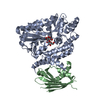 4xzvC 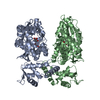 1hsjS S: Starting model for refinement C: citing same article ( |
|---|---|
| Similar structure data |

-Links
 PDBj
PDBj


- Assembly
| Deposited unit | 
| ||||||||||||||||||
|---|---|---|---|---|---|---|---|---|---|---|---|---|---|---|---|---|---|---|---|
| 1 | 
| ||||||||||||||||||
| 2 | 
| ||||||||||||||||||
| Unit cell |
| ||||||||||||||||||
| Noncrystallographic symmetry (NCS) | NCS domain:
NCS domain segments: Component-ID: 0 / Ens-ID: 1 / Beg auth comp-ID: LYS / Beg label comp-ID: LYS / End auth comp-ID: LYS / End label comp-ID: LYS / Refine code: 0 / Auth seq-ID: 1 - 419 / Label seq-ID: 2 - 420
|
-Components
| #1: Protein | Mass: 48939.320 Da / Num. of mol.: 2 Source method: isolated from a genetically manipulated source Source: (gene. exp.) Escherichia coli K-12 (bacteria), (gene. exp.) Homo sapiens (human)Gene: malE, b4034, JW3994, TRIAP1, 15E1.1, HSPC132 / Production host: Escherichia coli (E. coli) / References: UniProt: P0AEX9, UniProt: O43715#2: Polysaccharide |   , Oligosaccharide / Class: Nutrient / Mass: 342.297 Da / Num. of mol.: 2 , Oligosaccharide / Class: Nutrient / Mass: 342.297 Da / Num. of mol.: 2Source method: isolated from a genetically manipulated source Details: oligosaccharide / References: alpha-maltose #3: Water | ChemComp-HOH / | Water Mass: 18.015 Da / Num. of mol.: 266 / Source method: isolated from a natural source / Formula: H2O Mass: 18.015 Da / Num. of mol.: 266 / Source method: isolated from a natural source / Formula: H2O |
|---|
-Experimental details
-Experiment
| Experiment | Method: X-RAY DIFFRACTION |
|---|
- Sample preparation
| Crystal | Density Matthews: 2.08 Å3/Da / Density % sol: 40.74 % |
|---|---|
| Crystal grow | Temperature: 293 K / Method: vapor diffusion, sitting drop / pH: 4.6 Details: 100 mM sodium acetate, 25% (w/v) PEG 4000, 18% (w/v) MPD, 200 mM ammonium sulphate |
-Data collection
| Diffraction | Mean temperature: 100 K |
|---|---|
| Diffraction source | Source: SYNCHROTRON / Site: Diamond  / Beamline: I03 / Wavelength: 0.97949 Å / Beamline: I03 / Wavelength: 0.97949 Å |
| Detector | Type: PSI PILATUS 6M / Detector: PIXEL / Date: May 22, 2014 |
| Radiation | Protocol: SINGLE WAVELENGTH / Monochromatic (M) / Laue (L): M / Scattering type: x-ray |
| Radiation wavelength | Wavelength: 0.97949 Å / Relative weight: 1 |
| Reflection | Resolution: 2.12→48.41 Å / Num. obs: 45307 / % possible obs: 98.7 % / Redundancy: 3.6 % / Biso Wilson estimate: 35.5 Å2 / Rmerge(I) obs: 0.093 / Net I/σ(I): 9.3 |
| Reflection shell | Resolution: 2.12→2.18 Å / Redundancy: 3.2 % / Rmerge(I) obs: 0.585 / Mean I/σ(I) obs: 2.2 / % possible all: 94 |
- Processing
| Software |
| ||||||||||||||||||||||||||||||||||||||||||||||||||||||||||||||||||||||||||||||||||||||||||||||||||||||||||||||||||||||||||||||||||||||||||||||||||||||||||||||||||||||||||||||||||||||
|---|---|---|---|---|---|---|---|---|---|---|---|---|---|---|---|---|---|---|---|---|---|---|---|---|---|---|---|---|---|---|---|---|---|---|---|---|---|---|---|---|---|---|---|---|---|---|---|---|---|---|---|---|---|---|---|---|---|---|---|---|---|---|---|---|---|---|---|---|---|---|---|---|---|---|---|---|---|---|---|---|---|---|---|---|---|---|---|---|---|---|---|---|---|---|---|---|---|---|---|---|---|---|---|---|---|---|---|---|---|---|---|---|---|---|---|---|---|---|---|---|---|---|---|---|---|---|---|---|---|---|---|---|---|---|---|---|---|---|---|---|---|---|---|---|---|---|---|---|---|---|---|---|---|---|---|---|---|---|---|---|---|---|---|---|---|---|---|---|---|---|---|---|---|---|---|---|---|---|---|---|---|---|---|
| Refinement | Method to determine structure: MOLECULAR REPLACEMENT Starting model: 1hsj Resolution: 2.12→48.41 Å / Cor.coef. Fo:Fc: 0.961 / Cor.coef. Fo:Fc free: 0.939 / Cross valid method: THROUGHOUT / ESU R: 0.278 / ESU R Free: 0.208 / Stereochemistry target values: MAXIMUM LIKELIHOOD / Details: HYDROGENS HAVE BEEN ADDED IN THE RIDING POSITIONS
| ||||||||||||||||||||||||||||||||||||||||||||||||||||||||||||||||||||||||||||||||||||||||||||||||||||||||||||||||||||||||||||||||||||||||||||||||||||||||||||||||||||||||||||||||||||||
| Solvent computation | Ion probe radii: 0.8 Å / Shrinkage radii: 0.8 Å / VDW probe radii: 1.2 Å / Solvent model: MASK | ||||||||||||||||||||||||||||||||||||||||||||||||||||||||||||||||||||||||||||||||||||||||||||||||||||||||||||||||||||||||||||||||||||||||||||||||||||||||||||||||||||||||||||||||||||||
| Displacement parameters | Biso mean: 42.257 Å2
| ||||||||||||||||||||||||||||||||||||||||||||||||||||||||||||||||||||||||||||||||||||||||||||||||||||||||||||||||||||||||||||||||||||||||||||||||||||||||||||||||||||||||||||||||||||||
| Refinement step | Cycle: 1 / Resolution: 2.12→48.41 Å
| ||||||||||||||||||||||||||||||||||||||||||||||||||||||||||||||||||||||||||||||||||||||||||||||||||||||||||||||||||||||||||||||||||||||||||||||||||||||||||||||||||||||||||||||||||||||
| Refine LS restraints |
|