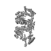
Entry Database : PDB / ID : 5j8qTitle Crystal Structure of the Cysteine Desulfurase SufS of Bacillus subtilis Cysteine desulfurase SufS Keywords / / / Function / homology Function Domain/homology Component
/ / / / / / / / / / / / / / / / / / / / / / / / Biological species Bacillus subtilis (bacteria)Method / / / Resolution : 1.702 Å Authors Altegoer, F. / Bange, G. Journal : Plos One / Year : 2016Title : Crystal Structure of Bacillus subtilis Cysteine Desulfurase SufS and Its Dynamic Interaction with Frataxin and Scaffold Protein SufU.Authors : Blauenburg, B. / Mielcarek, A. / Altegoer, F. / Fage, C.D. / Linne, U. / Bange, G. / Marahiel, M.A. History Deposition Apr 8, 2016 Deposition site / Processing site Revision 1.0 Jul 27, 2016 Provider / Type Revision 2.0 Jan 15, 2020 Group / Category / Item Revision 2.1 Jan 10, 2024 Group Data collection / Database references ... Data collection / Database references / Derived calculations / Refinement description Category chem_comp_atom / chem_comp_bond ... chem_comp_atom / chem_comp_bond / database_2 / pdbx_initial_refinement_model / struct_conn Item / _database_2.pdbx_database_accession / _struct_conn.pdbx_leaving_atom_flag
Show all Show less
 Movie
Movie Controller

 Yorodumi
 Open data
 Basic information
 Components
 Keywords
 TRANSFERASE /
  Function and homology information
 Authors
 Citation
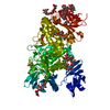
 Structure visualization
 Downloads & links
 Other downloads


 PDBj
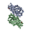
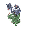
PDBj Assembly
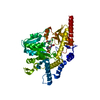


 Mass: 247.142 Da / Num. of mol.: 1 / Source method: obtained synthetically / Formula: C8H10NO6P
 Type: L-peptide linking / Mass: 89.093 Da / Num. of mol.: 1 / Source method: obtained synthetically / Formula: C3H7NO2
 Mass: 18.015 Da / Num. of mol.: 293 / Source method: isolated from a natural source / Formula: H2O
 Sample preparation
 / Beamline: ID29 / Wavelength: 0.972 Å
 Processing