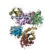
 Movie
Movie Controller


+ Open data

- Basic information
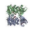
| Entry | Database: PDB / ID: 1hsj | ||||||
|---|---|---|---|---|---|---|---|
| Title | SARR MBP FUSION STRUCTURE | ||||||
 Components Components | FUSION PROTEIN CONSISTING OF STAPHYLOCOCCUS ACCESSORY REGULATOR PROTEIN R AND MALTOSE BINDING PROTEIN | ||||||
 Keywords Keywords | Transcription/Sugar Binding Protein / Novel fold for DNA binding / Transcription-Sugar Binding Protein COMPLEX | ||||||
| Function / homology |  Function and homology information Function and homology informationdetection of maltose stimulus / maltose binding / maltose transport complex / maltose transport / maltodextrin transmembrane transport / carbohydrate transmembrane transporter activity / ATP-binding cassette (ABC) transporter complex, substrate-binding subunit-containing / carbohydrate transport / : / ATP-binding cassette (ABC) transporter complex ...detection of maltose stimulus / maltose binding / maltose transport complex / maltose transport / maltodextrin transmembrane transport / carbohydrate transmembrane transporter activity / ATP-binding cassette (ABC) transporter complex, substrate-binding subunit-containing / carbohydrate transport / : / ATP-binding cassette (ABC) transporter complex / cell chemotaxis / outer membrane-bounded periplasmic space / periplasmic space / DNA damage response / regulation of DNA-templated transcription / DNA binding / membrane / cytoplasmSimilarity search - Function | ||||||
| Biological species |  Escherichia coli (E. coli) Escherichia coli (E. coli) Staphylococcus aureus (bacteria) Staphylococcus aureus (bacteria) | ||||||
| Method | X-RAY DIFFRACTION / SYNCHROTRON / MOLECULAR REPLACEMENT / Resolution: 2.3 Å | ||||||
 Authors Authors | Zhang, G. | ||||||
 Citation Citation | Journal: Proc.Natl.Acad.Sci.USA / Year: 2001 Title: Crystal structure of the SarR protein from Staphylococcus aureus. Authors: Liu, Y. / Manna, A. / Li, R. / Martin, W.E. / Murphy, R.C. / Cheung, A.L. / Zhang, G. | ||||||
| History |
|
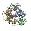
- Structure visualization
| Structure viewer | Molecule: MolmilJmol/JSmol |
|---|
- Downloads & links
-Download
| PDBx/mmCIF format | 1hsj.cif.gz | 197.9 KB | Display | PDBx/mmCIF format |
|---|---|---|---|---|
| PDB format | pdb1hsj.ent.gz | 159.2 KB | Display | PDB format |
| PDBx/mmJSON format | 1hsj.json.gz | Tree view | PDBx/mmJSON format | |
| Others |  Other downloads Other downloads |
-Validation report
| Arichive directory | https://data.pdbj.org/pub/pdb/validation_reports/hs/1hsjftp://data.pdbj.org/pub/pdb/validation_reports/hs/1hsj | HTTPS FTP |
|---|
-Related structure data
| Related structure data |  4mbpS S: Starting model for refinement |
|---|---|
| Similar structure data |


-Links
 PDBj
PDBj


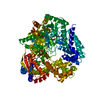
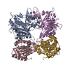
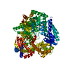
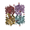
- Assembly
| Deposited unit | 
| ||||||||
|---|---|---|---|---|---|---|---|---|---|
| 1 |
| ||||||||
| Unit cell |
|
-Components
| #1: Protein | / SARR AND MBP / MMBP Mass: 54427.809 Da / Num. of mol.: 2 Source method: isolated from a genetically manipulated source Source: (gene. exp.) Escherichia coli (E. coli), (gene. exp.) Staphylococcus aureus (bacteria)Genus: Escherichia, Staphylococcus / Species: , / Production host: Escherichia coli (E. coli)References: UniProt: P0AEY0, UniProt: Q2YYV0, UniProt: P0AEX9*PLUS #2: Sugar | ChemComp-GLC / Glucose  Type: D-saccharide, alpha linking / Mass: 180.156 Da / Num. of mol.: 4 Type: D-saccharide, alpha linking / Mass: 180.156 Da / Num. of mol.: 4Source method: isolated from a genetically manipulated source Formula: C6H12O6 |
|---|
-Experimental details
-Experiment
| Experiment | Method: X-RAY DIFFRACTION / Number of used crystals: 1 |
|---|
- Sample preparation
| Crystal | Density Matthews: 2.59 Å3/Da / Density % sol: 52.59 % | ||||||||||||||||||||||||||||||||||||
|---|---|---|---|---|---|---|---|---|---|---|---|---|---|---|---|---|---|---|---|---|---|---|---|---|---|---|---|---|---|---|---|---|---|---|---|---|---|
| Crystal grow | *PLUS Method: vapor diffusion | ||||||||||||||||||||||||||||||||||||
| Components of the solutions | *PLUS
|
-Data collection
| Diffraction | Mean temperature: 95 K |
|---|---|
| Diffraction source | Source: SYNCHROTRON / Site: ALS  / Beamline: 5.0.2 / Wavelength: 0.99 / Beamline: 5.0.2 / Wavelength: 0.99 |
| Detector | Detector: CCD / Date: Nov 12, 2000 |
| Radiation | Protocol: SINGLE WAVELENGTH / Monochromatic (M) / Laue (L): M / Scattering type: x-ray |
| Radiation wavelength | Wavelength: 0.99 Å / Relative weight: 1 |
| Reflection | Resolution: 2.28→40 Å / Num. all: 50256 / Num. obs: 50256 / % possible obs: 89.9 % / Redundancy: 2 % / Biso Wilson estimate: 32 Å2 / Rmerge(I) obs: 0.028 / Rsym value: 0.032 / Net I/σ(I): 25.1 |
| Reflection shell | Resolution: 2.28→2.37 Å / Rmerge(I) obs: 0.449 / Mean I/σ(I) obs: 2.3 / % possible all: 84.8 |
| Reflection | *PLUS Num. obs: 37403 / % possible obs: 96.5 % / Num. measured all: 77545 |
- Processing
| Software |
| ||||||||||||||||||||
|---|---|---|---|---|---|---|---|---|---|---|---|---|---|---|---|---|---|---|---|---|---|
| Refinement | Method to determine structure: MOLECULAR REPLACEMENT Starting model: Maltose binding protein 4mbp Resolution: 2.3→19.95 Å / Rfactor Rfree error: 0.007 / Data cutoff high absF: 381018.15 / Data cutoff low absF: 0 / Isotropic thermal model: RESTRAINED / Cross valid method: THROUGHOUT / σ(F): 2
| ||||||||||||||||||||
| Solvent computation | Solvent model: FLAT MODEL / Bsol: 45.14 Å2 / ksol: 0.306 e/Å3 | ||||||||||||||||||||
| Displacement parameters | Biso mean: 71.3 Å2
| ||||||||||||||||||||
| Refine analyze |
| ||||||||||||||||||||
| Refinement step | Cycle: LAST / Resolution: 2.3→19.95 Å
| ||||||||||||||||||||
| Refine LS restraints |
| ||||||||||||||||||||
| LS refinement shell | Resolution: 2.3→2.44 Å / Rfactor Rfree error: 0.031 / Total num. of bins used: 6
| ||||||||||||||||||||
| Xplor file |
| ||||||||||||||||||||
| Software | *PLUS Name: CNS / Version: 0.9 / Classification: refinement | ||||||||||||||||||||
| Refinement | *PLUS Num. reflection obs: 37186 / σ(F): 2 / % reflection Rfree: 5 % / Rfactor obs: 0.2324 / Rfactor Rfree: 0.2822 | ||||||||||||||||||||
| Solvent computation | *PLUS | ||||||||||||||||||||
| Displacement parameters | *PLUS Biso mean: 71.3 Å2 | ||||||||||||||||||||
| Refine LS restraints | *PLUS
| ||||||||||||||||||||
| LS refinement shell | *PLUS Lowest resolution: 2.4 Å / Rfactor Rfree: 0.3802 / % reflection Rfree: 5 % / Rfactor Rwork: 0.404 / Num. reflection Rwork: 2702 / Rfactor obs: 0.4156 |
