 Movie
Movie Controller

[English] 日本語
 Yorodumi
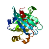
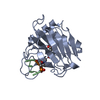
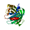
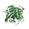
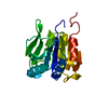
- PDB-4xw3: Crystal structure of the SPRY domain of the human DEAD-box protei... -
+ Open data

- Basic information
| Entry | Database: PDB / ID: 4xw3 | |||||||||
|---|---|---|---|---|---|---|---|---|---|---|
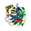
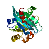
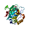
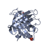
| Title | Crystal structure of the SPRY domain of the human DEAD-box protein DDX1 | |||||||||
 Components Components | ATP-dependent RNA helicase DDX1 | |||||||||
 Keywords Keywords |  HYDROLASE / beta-sandwich / insertion domain / DEAD-box protein / SPRY domain HYDROLASE / beta-sandwich / insertion domain / DEAD-box protein / SPRY domain | |||||||||
| Function / homology |  Function and homology information Function and homology informationcleavage body / tRNA-splicing ligase complex / DNA/RNA helicase activity / : / tRNA splicing, via endonucleolytic cleavage and ligation / protein localization to cytoplasmic stress granule / positive regulation of myeloid dendritic cell cytokine production / nuclease activity / poly(A) binding / tRNA processing in the nucleus ...cleavage body / tRNA-splicing ligase complex / DNA/RNA helicase activity / : / tRNA splicing, via endonucleolytic cleavage and ligation / protein localization to cytoplasmic stress granule / positive regulation of myeloid dendritic cell cytokine production / nuclease activity / poly(A) binding / tRNA processing in the nucleus / regulation of translational initiation / exonuclease activity / tRNA processing / DNA duplex unwinding / spliceosomal complex assembly / response to exogenous dsRNA / transcription coregulator activity / cytoplasmic stress granule / double-stranded RNA binding / double-strand break repair / positive regulation of canonical NF-kappaB signal transduction / defense response to virus / RNA helicase activity / RNA helicase / ribonucleoprotein complex / innate immune response / chromatin binding / ATP hydrolysis activity / mitochondrion / DNA binding / RNA binding / nucleoplasm / ATP binding / membrane / nucleus / cytosol / cytoplasmSimilarity search - Function | |||||||||
| Biological species |  Homo sapiens (human) Homo sapiens (human) | |||||||||
| Method | X-RAY DIFFRACTION / SYNCHROTRON / MOLECULAR REPLACEMENT / molecular replacement / Resolution: 2 Å | |||||||||
 Authors Authors | Kellner, J.N. / Meinhart, A. | |||||||||
| Funding support |  Germany, 1items Germany, 1items
| |||||||||
 Citation Citation | Journal: Acta Crystallogr.,Sect.F / Year: 2015 Title: Structure of the SPRY domain of the human RNA helicase DDX1, a putative interaction platform within a DEAD-box protein. Authors: Kellner, J.N. / Meinhart, A. | |||||||||
| History |
|
- Structure visualization
| Structure viewer | Molecule: MolmilJmol/JSmol |
|---|
- Downloads & links
-Download
| PDBx/mmCIF format | 4xw3.cif.gz | 164.8 KB | Display | PDBx/mmCIF format |
|---|---|---|---|---|
| PDB format | pdb4xw3.ent.gz | 129.1 KB | Display | PDB format |
| PDBx/mmJSON format | 4xw3.json.gz | Tree view | PDBx/mmJSON format | |
| Others |  Other downloads Other downloads |
-Validation report
| Arichive directory | https://data.pdbj.org/pub/pdb/validation_reports/xw/4xw3ftp://data.pdbj.org/pub/pdb/validation_reports/xw/4xw3 | HTTPS FTP |
|---|
-Related structure data
| Related structure data |  3tojS S: Starting model for refinement |
|---|---|
| Similar structure data |

-Links
 PDBj
PDBj


- Assembly
| Deposited unit | 
| ||||||||
|---|---|---|---|---|---|---|---|---|---|
| 1 | 
| ||||||||
| 2 | 
| ||||||||
| Unit cell |
| ||||||||
| Details | biological unit is the same as asym. |
-Components
| #1: Protein | Mass: 24644.887 Da / Num. of mol.: 2 / Fragment: unp residues 72-283 Source method: isolated from a genetically manipulated source Details: cDNA / Source: (gene. exp.) Homo sapiens (human) / Gene: DDX1, hCG_15914 / Plasmid: pET28a / Production host:  Escherichia coli BL21(DE3) (bacteria) / Strain (production host): CodonPlus- RIL / References: UniProt: A3RJH1, UniProt: Q92499*PLUS Escherichia coli BL21(DE3) (bacteria) / Strain (production host): CodonPlus- RIL / References: UniProt: A3RJH1, UniProt: Q92499*PLUS#2: Water | ChemComp-HOH / | Water Mass: 18.015 Da / Num. of mol.: 142 / Source method: isolated from a natural source / Formula: H2O Mass: 18.015 Da / Num. of mol.: 142 / Source method: isolated from a natural source / Formula: H2O |
|---|
-Experimental details
-Experiment
| Experiment | Method: X-RAY DIFFRACTION / Number of used crystals: 1 |
|---|
- Sample preparation
| Crystal | Density Matthews: 2.43 Å3/Da / Density % sol: 49.45 % / Description: spheroid crystals |
|---|---|
| Crystal grow | Temperature: 293 K / Method: vapor diffusion, hanging drop / pH: 5.5 Details: 35 % (v/v) PEG 600, 0.1 M tri-Sodium-citrate pH 5.5 - crystals appeared after 3 days and grew as single crystals PH range: 5 - 6 |
-Data collection
| Diffraction | Mean temperature: 100 K | ||||||||||||||||||||||||||||||||||||||||||||||||||||||||||||||||||||||||||||||||||||||||||||||||||||||||||||||||||||||||||||||||||||
|---|---|---|---|---|---|---|---|---|---|---|---|---|---|---|---|---|---|---|---|---|---|---|---|---|---|---|---|---|---|---|---|---|---|---|---|---|---|---|---|---|---|---|---|---|---|---|---|---|---|---|---|---|---|---|---|---|---|---|---|---|---|---|---|---|---|---|---|---|---|---|---|---|---|---|---|---|---|---|---|---|---|---|---|---|---|---|---|---|---|---|---|---|---|---|---|---|---|---|---|---|---|---|---|---|---|---|---|---|---|---|---|---|---|---|---|---|---|---|---|---|---|---|---|---|---|---|---|---|---|---|---|---|---|
| Diffraction source | Source: SYNCHROTRON / Site: SLS  / Beamline: X10SA / Wavelength: 1.06998 Å / Beamline: X10SA / Wavelength: 1.06998 Å | ||||||||||||||||||||||||||||||||||||||||||||||||||||||||||||||||||||||||||||||||||||||||||||||||||||||||||||||||||||||||||||||||||||
| Detector | Type: PSI PILATUS 6M / Detector: PIXEL / Date: Nov 10, 2012 | ||||||||||||||||||||||||||||||||||||||||||||||||||||||||||||||||||||||||||||||||||||||||||||||||||||||||||||||||||||||||||||||||||||
| Radiation | Monochromator: Si(111) / Protocol: SINGLE WAVELENGTH / Monochromatic (M) / Laue (L): M / Scattering type: x-ray | ||||||||||||||||||||||||||||||||||||||||||||||||||||||||||||||||||||||||||||||||||||||||||||||||||||||||||||||||||||||||||||||||||||
| Radiation wavelength | Wavelength: 1.06998 Å / Relative weight: 1 | ||||||||||||||||||||||||||||||||||||||||||||||||||||||||||||||||||||||||||||||||||||||||||||||||||||||||||||||||||||||||||||||||||||
| Reflection | Resolution: 2→47.78 Å / Num. obs: 28891 / % possible obs: 98.6 % / Observed criterion σ(I): -3 / Redundancy: 12.71 % / Biso Wilson estimate: 32.683 Å2 / Rmerge F obs: 0.074 / Rmerge(I) obs: 0.076 / Rrim(I) all: 0.079 / Χ2: 0.996 / Net I/σ(I): 23.63 / Num. measured all: 367329 | ||||||||||||||||||||||||||||||||||||||||||||||||||||||||||||||||||||||||||||||||||||||||||||||||||||||||||||||||||||||||||||||||||||
| Reflection shell | Diffraction-ID: 1 / Rejects: 0
|
-Phasing
| Phasing | Method: molecular replacement | |||||||||
|---|---|---|---|---|---|---|---|---|---|---|
| Phasing MR | Model details: Phaser MODE: MR_AUTO
|
- Processing
| Software |
| ||||||||||||||||||||||||||||||||||||||||||||||||||||||||||||||||||||||||||||||||||||||||||
|---|---|---|---|---|---|---|---|---|---|---|---|---|---|---|---|---|---|---|---|---|---|---|---|---|---|---|---|---|---|---|---|---|---|---|---|---|---|---|---|---|---|---|---|---|---|---|---|---|---|---|---|---|---|---|---|---|---|---|---|---|---|---|---|---|---|---|---|---|---|---|---|---|---|---|---|---|---|---|---|---|---|---|---|---|---|---|---|---|---|---|---|
| Refinement | Method to determine structure: MOLECULAR REPLACEMENT Starting model: 3toj Resolution: 2→47.78 Å / Cor.coef. Fo:Fc: 0.959 / Cor.coef. Fo:Fc free: 0.947 / SU B: 8.107 / SU ML: 0.113 / Cross valid method: THROUGHOUT / σ(F): 0 / ESU R: 0.187 / ESU R Free: 0.163 / Stereochemistry target values: MAXIMUM LIKELIHOOD / Details: HYDROGENS HAVE BEEN ADDED IN THE RIDING POSITIONS
| ||||||||||||||||||||||||||||||||||||||||||||||||||||||||||||||||||||||||||||||||||||||||||
| Solvent computation | Ion probe radii: 0.8 Å / Shrinkage radii: 0.8 Å / VDW probe radii: 1.2 Å / Solvent model: MASK | ||||||||||||||||||||||||||||||||||||||||||||||||||||||||||||||||||||||||||||||||||||||||||
| Displacement parameters | Biso max: 62.28 Å2 / Biso mean: 28.267 Å2 / Biso min: 12.98 Å2
| ||||||||||||||||||||||||||||||||||||||||||||||||||||||||||||||||||||||||||||||||||||||||||
| Refinement step | Cycle: final / Resolution: 2→47.78 Å
| ||||||||||||||||||||||||||||||||||||||||||||||||||||||||||||||||||||||||||||||||||||||||||
| Refine LS restraints |
| ||||||||||||||||||||||||||||||||||||||||||||||||||||||||||||||||||||||||||||||||||||||||||
| LS refinement shell | Resolution: 2→2.052 Å / Total num. of bins used: 20
| ||||||||||||||||||||||||||||||||||||||||||||||||||||||||||||||||||||||||||||||||||||||||||
| Refinement TLS params. | Method: refined / Refine-ID: X-RAY DIFFRACTION
| ||||||||||||||||||||||||||||||||||||||||||||||||||||||||||||||||||||||||||||||||||||||||||
| Refinement TLS group |
|
