 Movie
Movie Controller


+ Open data

- Basic information
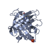
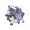
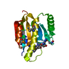
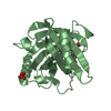
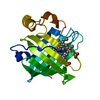
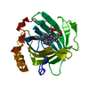
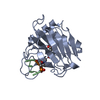
| Entry | Database: PDB / ID: 1chd | ||||||
|---|---|---|---|---|---|---|---|
| Title | CHEB METHYLESTERASE DOMAIN | ||||||
 Components Components | CHEB METHYLESTERASE | ||||||
 Keywords Keywords | CARBOXYL METHYLESTERASE /  CHEMOTAXIS PROTEIN / SERINE HYDROLASE CHEMOTAXIS PROTEIN / SERINE HYDROLASE | ||||||
| Function / homology |  Function and homology informationprotein-glutamate methylesterase / protein-glutamate methylesterase activity / protein-glutamine glutaminase activity / protein-glutamine glutaminase / phosphorelay response regulator activity / chemotaxis / cytoplasm Function and homology informationprotein-glutamate methylesterase / protein-glutamate methylesterase activity / protein-glutamine glutaminase activity / protein-glutamine glutaminase / phosphorelay response regulator activity / chemotaxis / cytoplasmSimilarity search - Function | ||||||
| Biological species |  Salmonella typhimurium (bacteria) Salmonella typhimurium (bacteria) | ||||||
| Method | X-RAY DIFFRACTION / Resolution: 1.75 Å | ||||||
 Authors Authors | West, A.H. / Martinez-Hackert, E. / Stock, A.M. | ||||||
 Citation Citation | Journal: J.Mol.Biol. / Year: 1995 Title: Crystal structure of the catalytic domain of the chemotaxis receptor methylesterase, CheB. Authors: West, A.H. / Martinez-Hackert, E. / Stock, A.M. #1: Journal: To be PublishedTitle: Purification, Crystallization, and Preliminary X-Ray Diffraction Analyses of the Bacterial Chemotaxis Receptor Modifying Enzymes Authors: West, A.H. / Djordjevic, S. / Martinez-Hackert, E. / Stock, A.M. #2: Journal: Biochim.Biophys.Acta / Year: 1992Title: Evidence that the Methylesterase of Bacterial Chemotaxis May be a Serine Hydrolase Authors: Krueger, J.K. / Stock, J. / Schutt, C.E. #3: Journal: Annu.Rev.Biophys.Biophys.Chem. / Year: 1991Title: Bacterial Chemotaxis and the Molecular Logic of Intracellular Signal Transduction Networks Authors: Stock, J.B. / Lukat, G.S. / Stock, A.M. #4: Journal: J.Biol.Chem. / Year: 1985Title: Multiple Forms of the Cheb Methylesterase in Bacterial Chemosensing Authors: Simms, S.A. / Keane, M.G. / Stock, J. | ||||||
| History |
| ||||||
| Remark 650 | HELIX HELIX DETERMINATION METHOD: H BONDS, DIHEDRAL ANGLES, KABSCH AND SANDER ALGORITHM, AND VISUAL ...HELIX HELIX DETERMINATION METHOD: H BONDS, DIHEDRAL ANGLES, KABSCH AND SANDER ALGORITHM, AND VISUAL INSPECTION HELIX_ID: L1,PART OF CONNECTING LOOP BETWEEN BETA STRAND 2 AND HELIX B. HELIX_ID: L2,PART OF CONNECTING LOOP BETWEEN BETA STRAND 6 AND HELIX C. HELIX_ID: L3,PART OF CONNECTING LOOP BETWEEN BETA STRAND 9 AND HELIX F. HELIX_ID: L4,PART OF CONNECTING LOOP BETWEEN BETA STRAND 8 AND HELIX E. | ||||||
| Remark 700 | SHEET SHEET SHEET_ID: SH1, H BONDS, DIHEDRAL ANGLES, KABSCH AND SANDER ALGORITHM, AND VISUAL ...SHEET SHEET SHEET_ID: SH1, H BONDS, DIHEDRAL ANGLES, KABSCH AND SANDER ALGORITHM, AND VISUAL INSPECTION. CENTRAL PARALLEL BETA SHEET. SHEET_ID: SH2, ANTIPARALLEL BETA MOTIF THAT FLANKS ONE SIDE OF THE CENTRAL SEVEN-STRANDED BETA SHEET. |
- Structure visualization
| Structure viewer | Molecule: MolmilJmol/JSmol |
|---|
- Downloads & links
-Download
| PDBx/mmCIF format | 1chd.cif.gz | 50.9 KB | Display | PDBx/mmCIF format |
|---|---|---|---|---|
| PDB format | pdb1chd.ent.gz | 36.2 KB | Display | PDB format |
| PDBx/mmJSON format | 1chd.json.gz | Tree view | PDBx/mmJSON format | |
| Others |  Other downloads Other downloads |
-Validation report
| Arichive directory | https://data.pdbj.org/pub/pdb/validation_reports/ch/1chdftp://data.pdbj.org/pub/pdb/validation_reports/ch/1chd | HTTPS FTP |
|---|
-Related structure data
| Similar structure data |
|---|


-Links
 PDBj
PDBj


- Assembly
| Deposited unit | 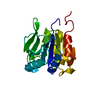
| ||||||||
|---|---|---|---|---|---|---|---|---|---|
| 1 |
| ||||||||
| Unit cell |
| ||||||||
| Atom site foot note | 1: CIS PROLINE - PRO 258 |
-Components
| #1: Protein | Mass: 21457.723 Da / Num. of mol.: 1 Source method: isolated from a genetically manipulated source Source: (gene. exp.) Salmonella typhimurium (bacteria) / Strain: LT2 / Gene: CHEB / Plasmid: PCP1 (PUC12 VECTOR) / Gene (production host): CHEB / Production host: Escherichia coli (E. coli) / Strain (production host): DH5ALPHAReferences: UniProt: P04042, protein-glutamate methylesterase | ||
|---|---|---|---|
| #2: Water | ChemComp-HOH / Water Mass: 18.015 Da / Num. of mol.: 123 / Source method: isolated from a natural source / Formula: H2O Mass: 18.015 Da / Num. of mol.: 123 / Source method: isolated from a natural source / Formula: H2O | ||
| Compound details | TURN TURN TYPES CLASSIFIED| Source details | MOLECULE_NAME: CHEB METHYLESTERASE DOMAIN. DETAILS OF CONSTRUCTION OF EXPRESSION PLASMID AND ...MOLECULE_NAME: CHEB METHYLESTE | |
-Experimental details
-Experiment
| Experiment | Method: X-RAY DIFFRACTION |
|---|
- Sample preparation
| Crystal | Density Matthews: 2.35 Å3/Da / Density % sol: 47.65 % | ||||||||||||||||||||||||||||||||||||
|---|---|---|---|---|---|---|---|---|---|---|---|---|---|---|---|---|---|---|---|---|---|---|---|---|---|---|---|---|---|---|---|---|---|---|---|---|---|
| Crystal grow | *PLUS pH: 8.5 / Method: vapor diffusion, hanging drop | ||||||||||||||||||||||||||||||||||||
| Components of the solutions | *PLUS
|
-Data collection
| Diffraction source | Wavelength: 1.5418 |
|---|---|
| Detector | Type: XUONG-HAMLIN MULTIWIRE / Detector: AREA DETECTOR / Date: Aug 16, 1994 |
| Radiation | Monochromatic (M) / Laue (L): M / Scattering type: x-ray |
| Radiation wavelength | Wavelength: 1.5418 Å / Relative weight: 1 |
| Reflection | Num. obs: 19729 / % possible obs: 93.6 % / Observed criterion σ(I): 3 / Redundancy: 2.4 % / Rmerge(I) obs: 0.045 |
| Reflection | *PLUS Highest resolution: 1.75 Å / Rmerge(I) obs: 0.045 |
| Reflection shell | *PLUS % possible obs: 89.3 % / Rmerge(I) obs: 0.154 / Mean I/σ(I) obs: 5 |
- Processing
| Software |
| ||||||||||||||||||||||||||||||||||||||||||||||||||||||||||||||||||||||||||||||||
|---|---|---|---|---|---|---|---|---|---|---|---|---|---|---|---|---|---|---|---|---|---|---|---|---|---|---|---|---|---|---|---|---|---|---|---|---|---|---|---|---|---|---|---|---|---|---|---|---|---|---|---|---|---|---|---|---|---|---|---|---|---|---|---|---|---|---|---|---|---|---|---|---|---|---|---|---|---|---|---|---|---|
| Refinement | Resolution: 1.75→8 Å / σ(F): 0 Details: UNUSUAL DIHEDRAL ANGLES (PHI= -140.17 AND PSI= -120.83) ARE OBSERVED FOR SER 164 WHICH MAY ACCOUNT FOR THE CLOSE CONTACT AS AN ACTIVE SITE RESIDUE AND IS PROPOSED TO BE INVOLVED IN THE ...Details: UNUSUAL DIHEDRAL ANGLES (PHI= -140.17 AND PSI= -120.83) ARE OBSERVED FOR SER 164 WHICH MAY ACCOUNT FOR THE CLOSE CONTACT AS AN ACTIVE SITE RESIDUE AND IS PROPOSED TO BE INVOLVED IN THE CATALYSIS OF METHYL ESTER HYDROLYSIS. PHI/PSI ANGLES THAT LIE OUTSIDE THE EXPECTED RANGE HAVE BEEN OBSERVED FOR MANY OTHER ACTIVE SITE SERINE NUCLEOPHILES IN HYDROLYTIC ENZYMES OF THIS CLASS.
| ||||||||||||||||||||||||||||||||||||||||||||||||||||||||||||||||||||||||||||||||
| Displacement parameters | Biso mean: 13.19 Å2 | ||||||||||||||||||||||||||||||||||||||||||||||||||||||||||||||||||||||||||||||||
| Refinement step | Cycle: LAST / Resolution: 1.75→8 Å
| ||||||||||||||||||||||||||||||||||||||||||||||||||||||||||||||||||||||||||||||||
| Refine LS restraints |
| ||||||||||||||||||||||||||||||||||||||||||||||||||||||||||||||||||||||||||||||||
| Software | *PLUS Name: ARP/PROLSQ/X-PLOR / Classification: refinement | ||||||||||||||||||||||||||||||||||||||||||||||||||||||||||||||||||||||||||||||||
| Refine LS restraints | *PLUS
|
