 Movie
Movie Controller

[English] 日本語
 Yorodumi
- PDB-4xuo: Structure of the CBM22-1 xylan-binding domain from Paenibacillus ... -
+ Open data

- Basic information
| Entry | Database: PDB / ID: 4xuo | ||||||
|---|---|---|---|---|---|---|---|
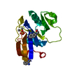
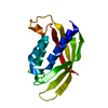
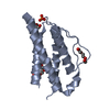
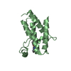
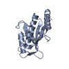
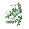
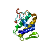
| Title | Structure of the CBM22-1 xylan-binding domain from Paenibacillus barcinonensis Xyn10C | ||||||
 Components Components | Endo-1,4-beta-xylanase C | ||||||
 Keywords Keywords | SUGAR BINDING PROTEIN /  Binding Site / Carbohydrates / Enzyme Stability / Substrate Specificity / Temperature / Endo-1 / 4-beta-xylanase / Xylan-binding domain / Calcium / Thermophilic enzymes / Thermostabilizing Domains Binding Site / Carbohydrates / Enzyme Stability / Substrate Specificity / Temperature / Endo-1 / 4-beta-xylanase / Xylan-binding domain / Calcium / Thermophilic enzymes / Thermostabilizing Domains | ||||||
| Function / homology |  Function and homology informationendo-1,4-beta-xylanase activity / endo-1,4-beta-xylanase / xylan catabolic process / carbohydrate binding Function and homology informationendo-1,4-beta-xylanase activity / endo-1,4-beta-xylanase / xylan catabolic process / carbohydrate bindingSimilarity search - Function | ||||||
| Biological species |  Paenibacillus barcinonensis (bacteria) Paenibacillus barcinonensis (bacteria) | ||||||
| Method | X-RAY DIFFRACTION / SYNCHROTRON / MOLECULAR REPLACEMENT / Resolution: 1.7 Å | ||||||
 Authors Authors | Sainz-Polo, M.A. / Sanz-Aparicio, J. | ||||||
 Citation Citation | Journal: J.Biol.Chem. / Year: 2015 Title: Exploring Multimodularity in Plant Cell Wall Deconstruction: STRUCTURAL AND FUNCTIONAL ANALYSIS OF Xyn10C CONTAINING THE CBM22-1-CBM22-2 TANDEM. Authors: Sainz-Polo, M.A. / Gonzalez, B. / Menendez, M. / Pastor, F.I. / Sanz-Aparicio, J. #1: Journal: Acta Crystallogr F Struct Biol Commun / Year: 2015 Title: Crystallization and preliminary X-ray diffraction analysis of the N-terminal domain of Paenibacillus barcinonensis xylanase 10C containing the CBM22-1-CBM22-2 tandem. Authors: Sainz-Polo, M.A. / Gonzalez, B. / Pastor, F.I. / Sanz-Aparicio, J. | ||||||
| History |
|
- Structure visualization
| Structure viewer | Molecule: MolmilJmol/JSmol |
|---|
- Downloads & links
-Download
| PDBx/mmCIF format | 4xuo.cif.gz | 74.4 KB | Display | PDBx/mmCIF format |
|---|---|---|---|---|
| PDB format | pdb4xuo.ent.gz | 54.8 KB | Display | PDB format |
| PDBx/mmJSON format | 4xuo.json.gz | Tree view | PDBx/mmJSON format | |
| Others |  Other downloads Other downloads |
-Validation report
| Arichive directory | https://data.pdbj.org/pub/pdb/validation_reports/xu/4xuoftp://data.pdbj.org/pub/pdb/validation_reports/xu/4xuo | HTTPS FTP |
|---|
-Related structure data
| Related structure data |  4w8lC  4xunC  4xupC  4xuqC  4xurC  4xutC  1dyoS S: Starting model for refinement C: citing same article ( |
|---|---|
| Similar structure data |


-Links
 PDBj
PDBj
- Assembly
| Deposited unit | 
| ||||||||||||||||||||||||||||||
|---|---|---|---|---|---|---|---|---|---|---|---|---|---|---|---|---|---|---|---|---|---|---|---|---|---|---|---|---|---|---|---|
| 1 | 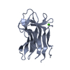
| ||||||||||||||||||||||||||||||
| 2 | 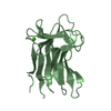
| ||||||||||||||||||||||||||||||
| Unit cell |
| ||||||||||||||||||||||||||||||
| Noncrystallographic symmetry (NCS) | NCS domain:
NCS domain segments: Component-ID: 1 / Ens-ID: 1 / Beg auth comp-ID: ALA / Beg label comp-ID: ALA / End auth comp-ID: ALA / End label comp-ID: ALA / Refine code: 6 / Auth seq-ID: 4 - 159 / Label seq-ID: 4 - 159
NCS oper:
|
-Components
| #1: Protein | Mass: 17294.076 Da / Num. of mol.: 2 / Fragment: UNP residues 29-186 Source method: isolated from a genetically manipulated source Source: (gene. exp.) Paenibacillus barcinonensis (bacteria) / Gene: xynC / Plasmid: pGEX-4T-2 / Production host: Escherichia coli BL21 (bacteria) / References: UniProt: O69230, endo-1,4-beta-xylanase#2: Chemical |   Mass: 40.078 Da / Num. of mol.: 2 / Source method: obtained synthetically / Formula: Ca Mass: 40.078 Da / Num. of mol.: 2 / Source method: obtained synthetically / Formula: Ca#3: Water | ChemComp-HOH / | Water Mass: 18.015 Da / Num. of mol.: 59 / Source method: isolated from a natural source / Formula: H2O Mass: 18.015 Da / Num. of mol.: 59 / Source method: isolated from a natural source / Formula: H2O |
|---|
-Experimental details
-Experiment
| Experiment | Method: X-RAY DIFFRACTION |
|---|
- Sample preparation
| Crystal | Density Matthews: 2.24 Å3/Da / Density % sol: 46.16 % |
|---|---|
| Crystal grow | Temperature: 293 K / Method: vapor diffusion, sitting drop Details: 0.2 M Sodium nitrate, 20% PEG 3350, ratio protein/precipitant: 1.5/1 |
-Data collection
| Diffraction | Mean temperature: 100 K |
|---|---|
| Diffraction source | Source: SYNCHROTRON / Site: ESRF  / Beamline: ID23-2 / Wavelength: 0.8729 Å / Beamline: ID23-2 / Wavelength: 0.8729 Å |
| Detector | Type: MARMOSAIC 225 mm CCD / Detector: CCD / Date: Sep 14, 2013 |
| Radiation | Monochromator: Horizontally side diffracting Silicon 111 crystal Protocol: SINGLE WAVELENGTH / Monochromatic (M) / Laue (L): M / Scattering type: x-ray |
| Radiation wavelength | Wavelength: 0.8729 Å / Relative weight: 1 |
| Reflection | Resolution: 1.7→30.54 Å / Num. obs: 32396 / % possible obs: 100 % / Observed criterion σ(F): 1 / Observed criterion σ(I): 1 / Redundancy: 5.7 % / Rmerge(I) obs: 0.135 / Rsym value: 0.062 / Net I/av σ(I): 9.7 / Net I/σ(I): 2.4 / Num. measured all: 184811 |
| Reflection shell | Resolution: 1.7→1.79 Å / Redundancy: 5.6 % / Rmerge(I) obs: 0.416 / Mean I/σ(I) obs: 1.7 / Num. unique all: 4730 / % possible all: 100 |
- Processing
| Software |
| ||||||||||||||||||||||||||||||||||||||||||||||||||||||||||||||||||||||||||||||||||||||||||||||||||||||||||||||||||||||||||||||||||||||||||||||||||||||||||||||||||||||||||||||||||||||
|---|---|---|---|---|---|---|---|---|---|---|---|---|---|---|---|---|---|---|---|---|---|---|---|---|---|---|---|---|---|---|---|---|---|---|---|---|---|---|---|---|---|---|---|---|---|---|---|---|---|---|---|---|---|---|---|---|---|---|---|---|---|---|---|---|---|---|---|---|---|---|---|---|---|---|---|---|---|---|---|---|---|---|---|---|---|---|---|---|---|---|---|---|---|---|---|---|---|---|---|---|---|---|---|---|---|---|---|---|---|---|---|---|---|---|---|---|---|---|---|---|---|---|---|---|---|---|---|---|---|---|---|---|---|---|---|---|---|---|---|---|---|---|---|---|---|---|---|---|---|---|---|---|---|---|---|---|---|---|---|---|---|---|---|---|---|---|---|---|---|---|---|---|---|---|---|---|---|---|---|---|---|---|---|
| Refinement | Method to determine structure: MOLECULAR REPLACEMENT Starting model: 1DYO Resolution: 1.7→30.54 Å / Cor.coef. Fo:Fc: 0.949 / Cor.coef. Fo:Fc free: 0.934 / SU B: 2.761 / SU ML: 0.092 / Cross valid method: THROUGHOUT / ESU R: 0.125 / ESU R Free: 0.121 / Stereochemistry target values: MAXIMUM LIKELIHOOD / Details: HYDROGENS HAVE BEEN ADDED IN THE RIDING POSITIONS
| ||||||||||||||||||||||||||||||||||||||||||||||||||||||||||||||||||||||||||||||||||||||||||||||||||||||||||||||||||||||||||||||||||||||||||||||||||||||||||||||||||||||||||||||||||||||
| Solvent computation | Ion probe radii: 0.8 Å / Shrinkage radii: 0.8 Å / VDW probe radii: 1.2 Å / Solvent model: MASK | ||||||||||||||||||||||||||||||||||||||||||||||||||||||||||||||||||||||||||||||||||||||||||||||||||||||||||||||||||||||||||||||||||||||||||||||||||||||||||||||||||||||||||||||||||||||
| Displacement parameters | Biso mean: 25.043 Å2
| ||||||||||||||||||||||||||||||||||||||||||||||||||||||||||||||||||||||||||||||||||||||||||||||||||||||||||||||||||||||||||||||||||||||||||||||||||||||||||||||||||||||||||||||||||||||
| Refinement step | Cycle: 1 / Resolution: 1.7→30.54 Å
| ||||||||||||||||||||||||||||||||||||||||||||||||||||||||||||||||||||||||||||||||||||||||||||||||||||||||||||||||||||||||||||||||||||||||||||||||||||||||||||||||||||||||||||||||||||||
| Refine LS restraints |
|