 Movie
Movie Controller

[English] 日本語
 Yorodumi
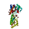
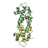
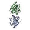
- PDB-4xih: Crystal structure of the R116A mutant AhpE from Mycobacterium tub... -
+ Open data

- Basic information
| Entry | Database: PDB / ID: 4xih | |||||||||
|---|---|---|---|---|---|---|---|---|---|---|
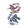
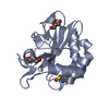
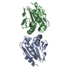
| Title | Crystal structure of the R116A mutant AhpE from Mycobacterium tuberculosis | |||||||||
 Components Components | AhpC/TSA family protein | |||||||||
 Keywords Keywords |  OXIDOREDUCTASE / Peroxiredoxins OXIDOREDUCTASE / Peroxiredoxins | |||||||||
| Function / homology |  Function and homology information Function and homology informationmycoredoxin-dependent peroxiredoxin / Tolerance by Mtb to nitric oxide produced by macrophages / response to nitrosative stress / peroxiredoxin activity / thioredoxin peroxidase activity / cell redox homeostasis / peroxidase activity / cellular response to oxidative stress / cytosol / cytoplasmSimilarity search - Function | |||||||||
| Biological species |   Mycobacterium tuberculosis (bacteria) Mycobacterium tuberculosis (bacteria) | |||||||||
| Method | X-RAY DIFFRACTION / SYNCHROTRON / MOLECULAR REPLACEMENT / molecular replacement / Resolution: 2.25 Å | |||||||||
 Authors Authors | Tamu Dufe, V. / van Molle, I. / Pallo, A. / Messens, J. | |||||||||
| Funding support |  Belgium, 1items Belgium, 1items
| |||||||||
 Citation Citation | Journal: Chem.Commun.(Camb.) / Year: 2016 Title: The active site architecture in peroxiredoxins: a case study on Mycobacterium tuberculosis AhpE. Authors: Pedre, B. / van Bergen, L.A. / Pallo, A. / Rosado, L.A. / Dufe, V.T. / Molle, I.V. / Wahni, K. / Erdogan, H. / Alonso, M. / Proft, F.D. / Messens, J. | |||||||||
| History |
|
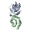
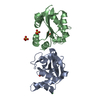
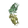
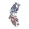
- Structure visualization
| Structure viewer | Molecule: MolmilJmol/JSmol |
|---|
- Downloads & links
-Download
| PDBx/mmCIF format | 4xih.cif.gz | 130 KB | Display | PDBx/mmCIF format |
|---|---|---|---|---|
| PDB format | pdb4xih.ent.gz | 102.1 KB | Display | PDB format |
| PDBx/mmJSON format | 4xih.json.gz | Tree view | PDBx/mmJSON format | |
| Others |  Other downloads Other downloads |
-Validation report
| Arichive directory | https://data.pdbj.org/pub/pdb/validation_reports/xi/4xihftp://data.pdbj.org/pub/pdb/validation_reports/xi/4xih | HTTPS FTP |
|---|
-Related structure data
| Related structure data |  5c04C  4x0xS S: Starting model for refinement C: citing same article ( |
|---|---|
| Similar structure data |


-Links
 PDBj
PDBj
- Assembly
| Deposited unit | 
| ||||||||
|---|---|---|---|---|---|---|---|---|---|
| 1 | 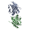
| ||||||||
| Unit cell |
|
-Components
| #1: Protein | Mass: 16814.902 Da / Num. of mol.: 2 / Mutation: R116A Source method: isolated from a genetically manipulated source Source: (gene. exp.) Mycobacterium tuberculosis (strain CCDC5079) (bacteria)Gene: ahpE, CCDC5079_2075, CFBS_2371 / Production host: Escherichia coli (E. coli) / References: UniProt: F7WEJ6, UniProt: P9WIE3*PLUS#2: Water | ChemComp-HOH / | Water Mass: 18.015 Da / Num. of mol.: 60 / Source method: isolated from a natural source / Formula: H2O Mass: 18.015 Da / Num. of mol.: 60 / Source method: isolated from a natural source / Formula: H2O |
|---|
-Experimental details
-Experiment
| Experiment | Method: X-RAY DIFFRACTION / Number of used crystals: 1 |
|---|
- Sample preparation
| Crystal | Density Matthews: 2.68 Å3/Da / Density % sol: 54.18 % |
|---|---|
| Crystal grow | Temperature: 293 K / Method: vapor diffusion, hanging drop / Details: 1.5 M Na-malonate, 0.1 M Na-acetate, pH 4.5 / PH range: 4.5 |
-Data collection
| Diffraction | Mean temperature: 100 K | ||||||||||||||||||||||||||||||||||||||||||||||||||||||||||||||||||||||||||||||||||||||||||||||||||||||||||||||||||||||||||||||||||||||||||||||||||||||||||||||||||||||||||||||||||||||||||||||||||||||||||||||||||
|---|---|---|---|---|---|---|---|---|---|---|---|---|---|---|---|---|---|---|---|---|---|---|---|---|---|---|---|---|---|---|---|---|---|---|---|---|---|---|---|---|---|---|---|---|---|---|---|---|---|---|---|---|---|---|---|---|---|---|---|---|---|---|---|---|---|---|---|---|---|---|---|---|---|---|---|---|---|---|---|---|---|---|---|---|---|---|---|---|---|---|---|---|---|---|---|---|---|---|---|---|---|---|---|---|---|---|---|---|---|---|---|---|---|---|---|---|---|---|---|---|---|---|---|---|---|---|---|---|---|---|---|---|---|---|---|---|---|---|---|---|---|---|---|---|---|---|---|---|---|---|---|---|---|---|---|---|---|---|---|---|---|---|---|---|---|---|---|---|---|---|---|---|---|---|---|---|---|---|---|---|---|---|---|---|---|---|---|---|---|---|---|---|---|---|---|---|---|---|---|---|---|---|---|---|---|---|---|---|---|---|---|
| Diffraction source | Source: SYNCHROTRON / Site: Diamond  / Beamline: I03 / Wavelength: 0.9763 Å / Beamline: I03 / Wavelength: 0.9763 Å | ||||||||||||||||||||||||||||||||||||||||||||||||||||||||||||||||||||||||||||||||||||||||||||||||||||||||||||||||||||||||||||||||||||||||||||||||||||||||||||||||||||||||||||||||||||||||||||||||||||||||||||||||||
| Detector | Type: DECTRIS PILATUS 6M / Detector: PIXEL / Date: Feb 23, 2013 | ||||||||||||||||||||||||||||||||||||||||||||||||||||||||||||||||||||||||||||||||||||||||||||||||||||||||||||||||||||||||||||||||||||||||||||||||||||||||||||||||||||||||||||||||||||||||||||||||||||||||||||||||||
| Radiation | Protocol: SINGLE WAVELENGTH / Monochromatic (M) / Laue (L): M / Scattering type: x-ray | ||||||||||||||||||||||||||||||||||||||||||||||||||||||||||||||||||||||||||||||||||||||||||||||||||||||||||||||||||||||||||||||||||||||||||||||||||||||||||||||||||||||||||||||||||||||||||||||||||||||||||||||||||
| Radiation wavelength | Wavelength: 0.9763 Å / Relative weight: 1 | ||||||||||||||||||||||||||||||||||||||||||||||||||||||||||||||||||||||||||||||||||||||||||||||||||||||||||||||||||||||||||||||||||||||||||||||||||||||||||||||||||||||||||||||||||||||||||||||||||||||||||||||||||
| Reflection | Resolution: 2.25→104 Å / Num. obs: 17069 / % possible obs: 97.5 % / Observed criterion σ(I): -3 / Redundancy: 2.73 % / Biso Wilson estimate: 45.05 Å2 / Rmerge F obs: 0.996 / Rmerge(I) obs: 0.098 / Rrim(I) all: 0.111 / Χ2: 1.009 / Net I/σ(I): 10.93 / Num. measured all: 74903 | ||||||||||||||||||||||||||||||||||||||||||||||||||||||||||||||||||||||||||||||||||||||||||||||||||||||||||||||||||||||||||||||||||||||||||||||||||||||||||||||||||||||||||||||||||||||||||||||||||||||||||||||||||
| Reflection shell | Diffraction-ID: 1 / Rejects: 0
|
-Phasing
| Phasing | Method: molecular replacement | |||||||||
|---|---|---|---|---|---|---|---|---|---|---|
| Phasing MR | Model details: Phaser MODE: MR_AUTO
|
- Processing
| Software |
| |||||||||||||||||||||||||||||||||||||||||||||||||||||||||||||||||||||||||||
|---|---|---|---|---|---|---|---|---|---|---|---|---|---|---|---|---|---|---|---|---|---|---|---|---|---|---|---|---|---|---|---|---|---|---|---|---|---|---|---|---|---|---|---|---|---|---|---|---|---|---|---|---|---|---|---|---|---|---|---|---|---|---|---|---|---|---|---|---|---|---|---|---|---|---|---|---|
| Refinement | Method to determine structure: MOLECULAR REPLACEMENT Starting model: 4X0X Resolution: 2.25→104 Å / Cor.coef. Fo:Fc: 0.945 / Cor.coef. Fo:Fc free: 0.936 / WRfactor Rfree: 0.2122 / WRfactor Rwork: 0.1927 / FOM work R set: 0.8058 / SU B: 14.934 / SU ML: 0.175 / SU R Cruickshank DPI: 0.3025 / SU Rfree: 0.2022 / Cross valid method: THROUGHOUT / σ(F): 0 / ESU R: 0.303 / ESU R Free: 0.202 / Stereochemistry target values: MAXIMUM LIKELIHOOD / Details: HYDROGENS HAVE BEEN ADDED IN THE RIDING POSITIONS
| |||||||||||||||||||||||||||||||||||||||||||||||||||||||||||||||||||||||||||
| Solvent computation | Ion probe radii: 0.8 Å / Shrinkage radii: 0.8 Å / VDW probe radii: 1.2 Å / Solvent model: MASK | |||||||||||||||||||||||||||||||||||||||||||||||||||||||||||||||||||||||||||
| Displacement parameters | Biso max: 91.87 Å2 / Biso mean: 40.799 Å2 / Biso min: 17.82 Å2
| |||||||||||||||||||||||||||||||||||||||||||||||||||||||||||||||||||||||||||
| Refinement step | Cycle: final / Resolution: 2.25→104 Å
| |||||||||||||||||||||||||||||||||||||||||||||||||||||||||||||||||||||||||||
| Refine LS restraints |
| |||||||||||||||||||||||||||||||||||||||||||||||||||||||||||||||||||||||||||
| LS refinement shell | Resolution: 2.25→2.309 Å
| |||||||||||||||||||||||||||||||||||||||||||||||||||||||||||||||||||||||||||
| Refinement TLS params. | Method: refined / Origin x: 24.5177 Å / Origin y: 121.1007 Å / Origin z: -35.7148 Å
| |||||||||||||||||||||||||||||||||||||||||||||||||||||||||||||||||||||||||||
| Refinement TLS group |
|
