 Movie
Movie Controller

[English] 日本語
 Yorodumi
- PDB-1xxu: Crystal Structure of AhpE from Mycrobacterium tuberculosis, a 1-C... -
+ Open data

- Basic information
| Entry | Database: PDB / ID: 1xxu | ||||||
|---|---|---|---|---|---|---|---|
| Title | Crystal Structure of AhpE from Mycrobacterium tuberculosis, a 1-Cys peroxiredoxin | ||||||
 Components Components | Hypothetical protein Rv2238c/MT2298 Hypothesis Hypothesis | ||||||
 Keywords Keywords | OXIDOREDUCTASE / 1-cys peroxiredoxin / thioredoxin fold / structural genomics / PSI / Protein Structure Initiative / TB Structural Genomics Consortium / TBSGC | ||||||
| Function / homology |  Function and homology information Function and homology informationmycoredoxin-dependent peroxiredoxin / Tolerance by Mtb to nitric oxide produced by macrophages / response to nitrosative stress / peroxiredoxin activity / thioredoxin peroxidase activity / cell redox homeostasis / peroxidase activity / cellular response to oxidative stress / cytosol / cytoplasmSimilarity search - Function | ||||||
| Biological species |   Mycobacterium tuberculosis (bacteria) Mycobacterium tuberculosis (bacteria) | ||||||
| Method | X-RAY DIFFRACTION / SYNCHROTRON / MOLECULAR REPLACEMENT / Resolution: 1.9 Å | ||||||
 Authors Authors | Li, S. / Peterson, N.A. / Kim, M.Y. / Kim, C.Y. / Hung, L.W. / Yu, M. / Lekin, T. / Segelke, B.W. / Lott, J.S. / Baker, E.N. / TB Structural Genomics Consortium (TBSGC) | ||||||
 Citation Citation | Journal: J.Mol.Biol. / Year: 2005 Title: Crystal Structure of AhpE from Mycobacterium tuberculosis, a 1-Cys Peroxiredoxin Authors: Li, S. / Peterson, N.A. / Kim, M.Y. / Kim, C.Y. / Hung, L.W. / Yu, M. / Lekin, T. / Segelke, B.W. / Lott, J.S. / Baker, E.N. | ||||||
| History |
|
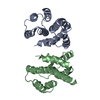
- Structure visualization
| Structure viewer | Molecule: MolmilJmol/JSmol |
|---|
- Downloads & links
-Download
| PDBx/mmCIF format | 1xxu.cif.gz | 128 KB | Display | PDBx/mmCIF format |
|---|---|---|---|---|
| PDB format | pdb1xxu.ent.gz | 101.7 KB | Display | PDB format |
| PDBx/mmJSON format | 1xxu.json.gz | Tree view | PDBx/mmJSON format | |
| Others |  Other downloads Other downloads |
-Validation report
| Arichive directory | https://data.pdbj.org/pub/pdb/validation_reports/xx/1xxuftp://data.pdbj.org/pub/pdb/validation_reports/xx/1xxu | HTTPS FTP |
|---|
-Related structure data
| Related structure data |  1xvwSC S: Starting model for refinement C: citing same article ( |
|---|---|
| Similar structure data | |
| Other databases |


-Links
 PDBj
PDBj
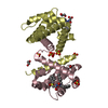
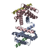
- Assembly
| Deposited unit | 
| ||||||||
|---|---|---|---|---|---|---|---|---|---|
| 1 | 
| ||||||||
| 2 | 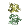
| ||||||||
| Unit cell |
| ||||||||
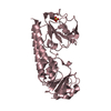
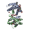
| Details | The biological assembly is a octamer generated from crystallographic 4-fold axis |
-Components
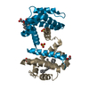
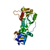
| #1: Protein | Hypothesis / 1-Cys peroxiredoxin Mass: 16833.949 Da / Num. of mol.: 4 Source method: isolated from a genetically manipulated source Source: (gene. exp.) Mycobacterium tuberculosis (bacteria) / Gene: Rv2238c / Plasmid: pProEX HTa / Production host: Escherichia coli (E. coli) / Strain (production host): BL21(DE3)pRIReferences: UniProt: P65688, UniProt: P9WIE3*PLUS, peroxidase#2: Water | ChemComp-HOH / | Water Mass: 18.015 Da / Num. of mol.: 188 / Source method: isolated from a natural source / Formula: H2O Mass: 18.015 Da / Num. of mol.: 188 / Source method: isolated from a natural source / Formula: H2O |
|---|
-Experimental details
-Experiment
| Experiment | Method: X-RAY DIFFRACTION / Number of used crystals: 1 |
|---|
- Sample preparation
| Crystal | Density Matthews: 2.8 Å3/Da / Density % sol: 55.1 % |
|---|---|
| Crystal grow | Temperature: 291 K / Method: vapor diffusion, sitting drop / pH: 4.5 Details: 1.8M sodium malonate pH 5.0 mixed with 0.1M sodium acetate pH 4.5, VAPOR DIFFUSION, SITTING DROP, temperature 291K |
-Data collection
| Diffraction | Mean temperature: 113 K |
|---|---|
| Diffraction source | Source: SYNCHROTRON / Site: ALS  / Beamline: 5.0.2 / Wavelength: 0.91983 Å / Beamline: 5.0.2 / Wavelength: 0.91983 Å |
| Detector | Type: ADSC QUANTUM 210 / Detector: CCD |
| Radiation | Protocol: SINGLE WAVELENGTH / Monochromatic (M) / Laue (L): M / Scattering type: x-ray |
| Radiation wavelength | Wavelength: 0.91983 Å / Relative weight: 1 |
| Reflection | Resolution: 1.9→37.05 Å / Num. all: 59088 / Num. obs: 59088 / % possible obs: 99.8 % / Observed criterion σ(F): 0 / Biso Wilson estimate: 15.7 Å2 / Rsym value: 0.082 / Net I/σ(I): 16.2 |
| Reflection shell | Resolution: 1.9→1.97 Å / Redundancy: 99.6 % / Mean I/σ(I) obs: 2.29 / Num. unique all: 5817 / Rsym value: 0.598 |
- Processing
| Software |
| |||||||||||||||||||||||||||||||||||||||||||||||||
|---|---|---|---|---|---|---|---|---|---|---|---|---|---|---|---|---|---|---|---|---|---|---|---|---|---|---|---|---|---|---|---|---|---|---|---|---|---|---|---|---|---|---|---|---|---|---|---|---|---|---|
| Refinement | Method to determine structure: MOLECULAR REPLACEMENT Starting model: 1xvw Resolution: 1.9→37.05 Å / Cross valid method: THROUGHOUT / Details: CNS bulk solvent model used
| |||||||||||||||||||||||||||||||||||||||||||||||||
| Displacement parameters | Biso mean: 22.25 Å2
| |||||||||||||||||||||||||||||||||||||||||||||||||
| Refine analyze |
| |||||||||||||||||||||||||||||||||||||||||||||||||
| Refinement step | Cycle: LAST / Resolution: 1.9→37.05 Å
| |||||||||||||||||||||||||||||||||||||||||||||||||
| Refine LS restraints |
| |||||||||||||||||||||||||||||||||||||||||||||||||
| LS refinement shell | Refine-ID: X-RAY DIFFRACTION / Total num. of bins used: 6
|
