 Movie
Movie Controller

[English] 日本語
 Yorodumi
- PDB-1xvw: Crystal Structure of AhpE from Mycobacterium tuberculosis, a 1-Cy... -
+ Open data

- Basic information
| Entry | Database: PDB / ID: 1xvw | ||||||
|---|---|---|---|---|---|---|---|
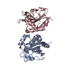
| Title | Crystal Structure of AhpE from Mycobacterium tuberculosis, a 1-Cys peroxiredoxin | ||||||
 Components Components | Hypothetical protein Rv2238c/MT2298 Hypothesis Hypothesis | ||||||
 Keywords Keywords | OXIDOREDUCTASE / thioredoxin fold / Oxidized cystein sulfenic acid / Structural Genomics / PSI / Protein Structure Initiative / TB Structural Genomics Consortium / TBSGC | ||||||
| Function / homology |  Function and homology information Function and homology informationmycoredoxin-dependent peroxiredoxin / Tolerance by Mtb to nitric oxide produced by macrophages / response to nitrosative stress / peroxiredoxin activity / thioredoxin peroxidase activity / cell redox homeostasis / peroxidase activity / cellular response to oxidative stress / cytosol / cytoplasmSimilarity search - Function | ||||||
| Biological species |   Mycobacterium tuberculosis (bacteria) Mycobacterium tuberculosis (bacteria) | ||||||
| Method | X-RAY DIFFRACTION / SYNCHROTRON / MAD / Resolution: 1.9 Å | ||||||
 Authors Authors | Li, S. / Peterson, N.A. / Kim, M.Y. / Kim, C.Y. / Hung, L.W. / Yu, M. / Lekin, T. / Segelke, B.W. / Lott, J.S. / Baker, E.N. / TB Structural Genomics Consortium (TBSGC) | ||||||
 Citation Citation | Journal: J.Mol.Biol. / Year: 2005 Title: Crystal Structure of AhpE from Mycobacterium tuberculosis, a 1-Cys Peroxiredoxin Authors: Li, S. / Peterson, N.A. / Kim, M.Y. / Kim, C.Y. / Hung, L.W. / Yu, M. / Lekin, T. / Segelke, B.W. / Lott, J.S. / Baker, E.N. | ||||||
| History |
|
- Structure visualization
| Structure viewer | Molecule: MolmilJmol/JSmol |
|---|
- Downloads & links
-Download
| PDBx/mmCIF format | 1xvw.cif.gz | 72.8 KB | Display | PDBx/mmCIF format |
|---|---|---|---|---|
| PDB format | pdb1xvw.ent.gz | 58.6 KB | Display | PDB format |
| PDBx/mmJSON format | 1xvw.json.gz | Tree view | PDBx/mmJSON format | |
| Others |  Other downloads Other downloads |
-Validation report
| Arichive directory | https://data.pdbj.org/pub/pdb/validation_reports/xv/1xvwftp://data.pdbj.org/pub/pdb/validation_reports/xv/1xvw | HTTPS FTP |
|---|
-Related structure data


-Links
 PDBj
PDBj
- Assembly
| Deposited unit | 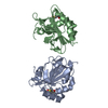
| ||||||||
|---|---|---|---|---|---|---|---|---|---|
| 1 | 
| ||||||||
| Unit cell |
| ||||||||
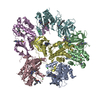
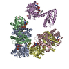
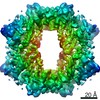
| Details | The biological assembly is a octamer generated from the dimer in the asymmetric unit by the crystallographic 4-fold symmetry |
-Components
| #1: Protein | Hypothesis / 1-cys alkylhydroperoxiredoxin reductase Mass: 17614.850 Da / Num. of mol.: 2 Source method: isolated from a genetically manipulated source Source: (gene. exp.) Mycobacterium tuberculosis (bacteria) / Gene: rv2238c / Plasmid: pProEX HTa / Species (production host): Escherichia coli / Production host: Escherichia coli BL21(DE3) (bacteria) / Strain (production host): BL21(DE3)References: UniProt: P65688, UniProt: P9WIE3*PLUS, Oxidoreductases; Acting on a sulfur group of donors; With NAD+ or NADP+ as acceptor#2: Water | ChemComp-HOH / | Water Mass: 18.015 Da / Num. of mol.: 184 / Source method: isolated from a natural source / Formula: H2O Mass: 18.015 Da / Num. of mol.: 184 / Source method: isolated from a natural source / Formula: H2O |
|---|
-Experimental details
-Experiment
| Experiment | Method: X-RAY DIFFRACTION / Number of used crystals: 1 |
|---|
- Sample preparation
| Crystal | Density Matthews: 4.61 Å3/Da / Density % sol: 47 % |
|---|---|
| Crystal grow | Temperature: 291 K / Method: vapor diffusion, sitting drop / pH: 4.5 Details: 1.8M sodium malonate pH 5.0, 0.1M sodium acetate pH 4.5, VAPOR DIFFUSION, SITTING DROP, temperature 291K |
-Data collection
| Diffraction | Mean temperature: 113 K | |||||||||||||||
|---|---|---|---|---|---|---|---|---|---|---|---|---|---|---|---|---|
| Diffraction source | Source: SYNCHROTRON / Site: ALS  / Beamline: 5.0.2 / Wavelength: 1.00000, 0.97937, 0.97918, 0.95370 / Beamline: 5.0.2 / Wavelength: 1.00000, 0.97937, 0.97918, 0.95370 | |||||||||||||||
| Detector | Type: ADSC QUANTUM 210 / Detector: CCD | |||||||||||||||
| Radiation | Protocol: MAD / Monochromatic (M) / Laue (L): M / Scattering type: x-ray | |||||||||||||||
| Radiation wavelength |
| |||||||||||||||
| Reflection | Resolution: 1.87→46.8 Å / Num. obs: 33679 / Redundancy: 97.6 % / Biso Wilson estimate: 17.4 Å2 / Net I/σ(I): 17.9 | |||||||||||||||
| Reflection shell | Resolution: 1.8→1.86 Å / Redundancy: 80.7 % / Mean I/σ(I) obs: 1.58 / Num. unique all: 2764 |
- Processing
| Software |
| |||||||||||||||||||||||||||||||||||||||||||||||||
|---|---|---|---|---|---|---|---|---|---|---|---|---|---|---|---|---|---|---|---|---|---|---|---|---|---|---|---|---|---|---|---|---|---|---|---|---|---|---|---|---|---|---|---|---|---|---|---|---|---|---|
| Refinement | Method to determine structure: MAD / Resolution: 1.9→46.8 Å / Cross valid method: THROUGHOUT / Stereochemistry target values: Engh & Huber
| |||||||||||||||||||||||||||||||||||||||||||||||||
| Displacement parameters |
| |||||||||||||||||||||||||||||||||||||||||||||||||
| Refine analyze |
| |||||||||||||||||||||||||||||||||||||||||||||||||
| Refinement step | Cycle: LAST / Resolution: 1.9→46.8 Å
| |||||||||||||||||||||||||||||||||||||||||||||||||
| Refine LS restraints |
| |||||||||||||||||||||||||||||||||||||||||||||||||
| LS refinement shell | Refine-ID: X-RAY DIFFRACTION / Total num. of bins used: 6
|
