 Movie
Movie Controller

[English] 日本語
 Yorodumi
- PDB-4wn5: Crystal structure of the C-terminal Per-Arnt-Sim (PASb) of human ... -
+ Open data

- Basic information
| Entry | Database: PDB / ID: 4wn5 | |||||||||||||||
|---|---|---|---|---|---|---|---|---|---|---|---|---|---|---|---|---|
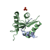
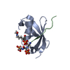
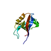
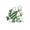
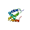
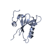
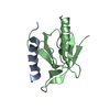
| Title | Crystal structure of the C-terminal Per-Arnt-Sim (PASb) of human HIF-3alpha9 bound to 18:1-1-monoacylglycerol | |||||||||||||||
 Components Components | Hypoxia-inducible factor 3-alpha | |||||||||||||||
 Keywords Keywords |  TRANSCRIPTION / HIF-3alpha / PAS domain / monoacylglycerol / lipid / fatty acid TRANSCRIPTION / HIF-3alpha / PAS domain / monoacylglycerol / lipid / fatty acid | |||||||||||||||
| Function / homology |  Function and homology information: / Transcriptional regulation of pluripotent stem cells / chromatin => GO:0000785 / Regulation of gene expression by Hypoxia-inducible Factor / post-translational protein modification / Oxygen-dependent proline hydroxylation of Hypoxia-inducible Factor Alpha / Neddylation / angiogenesis / transcription by RNA polymerase II / protein ubiquitination ...: / Transcriptional regulation of pluripotent stem cells / chromatin => GO:0000785 / Regulation of gene expression by Hypoxia-inducible Factor / post-translational protein modification / Oxygen-dependent proline hydroxylation of Hypoxia-inducible Factor Alpha / Neddylation / angiogenesis / transcription by RNA polymerase II / protein ubiquitination / protein dimerization activity / DNA-binding transcription factor activity, RNA polymerase II-specific / nuclear speck / RNA polymerase II cis-regulatory region sequence-specific DNA binding / apoptotic process / regulation of transcription by RNA polymerase II / positive regulation of transcription by RNA polymerase II / mitochondrion / nucleoplasm / nucleus / plasma membrane / cytosol Function and homology information: / Transcriptional regulation of pluripotent stem cells / chromatin => GO:0000785 / Regulation of gene expression by Hypoxia-inducible Factor / post-translational protein modification / Oxygen-dependent proline hydroxylation of Hypoxia-inducible Factor Alpha / Neddylation / angiogenesis / transcription by RNA polymerase II / protein ubiquitination ...: / Transcriptional regulation of pluripotent stem cells / chromatin => GO:0000785 / Regulation of gene expression by Hypoxia-inducible Factor / post-translational protein modification / Oxygen-dependent proline hydroxylation of Hypoxia-inducible Factor Alpha / Neddylation / angiogenesis / transcription by RNA polymerase II / protein ubiquitination / protein dimerization activity / DNA-binding transcription factor activity, RNA polymerase II-specific / nuclear speck / RNA polymerase II cis-regulatory region sequence-specific DNA binding / apoptotic process / regulation of transcription by RNA polymerase II / positive regulation of transcription by RNA polymerase II / mitochondrion / nucleoplasm / nucleus / plasma membrane / cytosolSimilarity search - Function | |||||||||||||||
| Biological species |  Homo sapiens (human) Homo sapiens (human) | |||||||||||||||
| Method | X-RAY DIFFRACTION / SYNCHROTRON / MOLECULAR REPLACEMENT / Resolution: 1.15 Å | |||||||||||||||
 Authors Authors | Fala, A.M. / Oliveira, J.F. / Dias, S.M. / Ambrosio, A.L. | |||||||||||||||
| Funding support |  Brazil, 4items Brazil, 4items
| |||||||||||||||
 Citation Citation | Journal: Sci Rep / Year: 2015 Title: Unsaturated fatty acids as high-affinity ligands of the C-terminal Per-ARNT-Sim domain from the Hypoxia-inducible factor 3 alpha. Authors: Fala, A.M. / Oliveira, J.F. / Adamoski, D. / Aricetti, J.A. / Dias, M.M. / Dias, M.V.B. / Sforca, M.L. / Lopes-de-Oliveira, P.S. / Rocco, S.A. / Caldana, C. / Dias, S.M.G. / Ambrosio, A.L.B. | |||||||||||||||
| History |
|
- Structure visualization
| Structure viewer | Molecule: MolmilJmol/JSmol |
|---|
- Downloads & links
-Download
| PDBx/mmCIF format | 4wn5.cif.gz | 157.1 KB | Display | PDBx/mmCIF format |
|---|---|---|---|---|
| PDB format | pdb4wn5.ent.gz | 126.9 KB | Display | PDB format |
| PDBx/mmJSON format | 4wn5.json.gz | Tree view | PDBx/mmJSON format | |
| Others |  Other downloads Other downloads |
-Validation report
| Arichive directory | https://data.pdbj.org/pub/pdb/validation_reports/wn/4wn5ftp://data.pdbj.org/pub/pdb/validation_reports/wn/4wn5 | HTTPS FTP |
|---|
-Related structure data
| Related structure data | 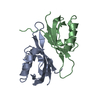 4h6jS S: Starting model for refinement |
|---|---|
| Similar structure data |


-Links
 PDBj
PDBj


- Assembly
| Deposited unit | 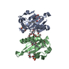
| ||||||||||||
|---|---|---|---|---|---|---|---|---|---|---|---|---|---|
| 1 | 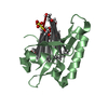
| ||||||||||||
| 2 | 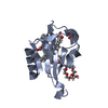
| ||||||||||||
| Unit cell |
| ||||||||||||
| Components on special symmetry positions |
|
-Components
| #1: Protein | Mass: 12520.955 Da / Num. of mol.: 2 / Fragment: unp residues 235-345 Source method: isolated from a genetically manipulated source Source: (gene. exp.) Homo sapiens (human) / Gene: HIF3A, BHLHE17, MOP7, PASD7 / Plasmid: pET28a / Production host:  Escherichia coli (E. coli) / Strain (production host): Rosetta-2 / References: UniProt: Q9Y2N7 Escherichia coli (E. coli) / Strain (production host): Rosetta-2 / References: UniProt: Q9Y2N7#2: Chemical | Sulfate  Mass: 96.063 Da / Num. of mol.: 2 / Source method: obtained synthetically / Formula: SO4 Mass: 96.063 Da / Num. of mol.: 2 / Source method: obtained synthetically / Formula: SO4#3: Chemical |   Mass: 356.540 Da / Num. of mol.: 2 / Source method: obtained synthetically / Formula: C21H40O4 Mass: 356.540 Da / Num. of mol.: 2 / Source method: obtained synthetically / Formula: C21H40O4#4: Chemical | ChemComp-P6G / Polyethylene glycol  Mass: 282.331 Da / Num. of mol.: 5 / Source method: obtained synthetically / Formula: C12H26O7 / Comment: precipitant*YM Mass: 282.331 Da / Num. of mol.: 5 / Source method: obtained synthetically / Formula: C12H26O7 / Comment: precipitant*YM#5: Water | ChemComp-HOH / | Water Mass: 18.015 Da / Num. of mol.: 295 / Source method: isolated from a natural source / Formula: H2O Mass: 18.015 Da / Num. of mol.: 295 / Source method: isolated from a natural source / Formula: H2O |
|---|
-Experimental details
-Experiment
| Experiment | Method: X-RAY DIFFRACTION |
|---|
- Sample preparation
| Crystal | Density Matthews: 1.92 Å3/Da / Density % sol: 36.06 % |
|---|---|
| Crystal grow | Temperature: 291 K / Method: vapor diffusion, sitting drop / pH: 5.9 Details: 3% PEG 400, 1.95 M ammonium sulfate, 0.1 M sodium acetate |
-Data collection
| Diffraction | Mean temperature: 100 K |
|---|---|
| Diffraction source | Source: SYNCHROTRON / Site: PETRA III, EMBL c/o DESY  / Beamline: P13 (MX1) / Wavelength: 0.97724 Å / Beamline: P13 (MX1) / Wavelength: 0.97724 Å |
| Detector | Type: DECTRIS PILATUS 6M-F / Detector: PIXEL / Date: Sep 25, 2013 |
| Radiation | Protocol: SINGLE WAVELENGTH / Monochromatic (M) / Laue (L): M / Scattering type: x-ray |
| Radiation wavelength | Wavelength: 0.97724 Å / Relative weight: 1 |
| Reflection | Resolution: 1.15→28.5 Å / Num. obs: 68827 / % possible obs: 99.6 % / Redundancy: 9.8 % / Net I/σ(I): 11.5 |
| Reflection shell | Resolution: 1.15→1.17 Å / Redundancy: 9.1 % / % possible all: 99.4 |
- Processing
| Software | Name: PHENIX / Version: (phenix.refine: 1.8.4_1496) / Classification: refinement | |||||||||||||||||||||||||||||||||||||||||||||||||||||||||||||||||||||||||||||||||||||||||||||||||||||||||||||||||||||||||||||||||||||||||||||||||||||||||||||||||||||||||||||||||||||||||||||||||||||||||||||||||||||||||
|---|---|---|---|---|---|---|---|---|---|---|---|---|---|---|---|---|---|---|---|---|---|---|---|---|---|---|---|---|---|---|---|---|---|---|---|---|---|---|---|---|---|---|---|---|---|---|---|---|---|---|---|---|---|---|---|---|---|---|---|---|---|---|---|---|---|---|---|---|---|---|---|---|---|---|---|---|---|---|---|---|---|---|---|---|---|---|---|---|---|---|---|---|---|---|---|---|---|---|---|---|---|---|---|---|---|---|---|---|---|---|---|---|---|---|---|---|---|---|---|---|---|---|---|---|---|---|---|---|---|---|---|---|---|---|---|---|---|---|---|---|---|---|---|---|---|---|---|---|---|---|---|---|---|---|---|---|---|---|---|---|---|---|---|---|---|---|---|---|---|---|---|---|---|---|---|---|---|---|---|---|---|---|---|---|---|---|---|---|---|---|---|---|---|---|---|---|---|---|---|---|---|---|---|---|---|---|---|---|---|---|---|---|---|---|---|---|---|---|
| Refinement | Method to determine structure: MOLECULAR REPLACEMENT Starting model: 4h6j Resolution: 1.15→28.5 Å / SU ML: 0.06 / Cross valid method: FREE R-VALUE / σ(F): 1.91 / Phase error: 10.42 / Stereochemistry target values: ML
| |||||||||||||||||||||||||||||||||||||||||||||||||||||||||||||||||||||||||||||||||||||||||||||||||||||||||||||||||||||||||||||||||||||||||||||||||||||||||||||||||||||||||||||||||||||||||||||||||||||||||||||||||||||||||
| Solvent computation | Shrinkage radii: 0.9 Å / VDW probe radii: 1.11 Å / Solvent model: FLAT BULK SOLVENT MODEL | |||||||||||||||||||||||||||||||||||||||||||||||||||||||||||||||||||||||||||||||||||||||||||||||||||||||||||||||||||||||||||||||||||||||||||||||||||||||||||||||||||||||||||||||||||||||||||||||||||||||||||||||||||||||||
| Refinement step | Cycle: LAST / Resolution: 1.15→28.5 Å
| |||||||||||||||||||||||||||||||||||||||||||||||||||||||||||||||||||||||||||||||||||||||||||||||||||||||||||||||||||||||||||||||||||||||||||||||||||||||||||||||||||||||||||||||||||||||||||||||||||||||||||||||||||||||||
| Refine LS restraints |
| |||||||||||||||||||||||||||||||||||||||||||||||||||||||||||||||||||||||||||||||||||||||||||||||||||||||||||||||||||||||||||||||||||||||||||||||||||||||||||||||||||||||||||||||||||||||||||||||||||||||||||||||||||||||||
| LS refinement shell |
|
