 Movie
Movie Controller


+ Open data

- Basic information
| Entry | Database: PDB / ID: 3fg7 | ||||||
|---|---|---|---|---|---|---|---|
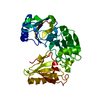
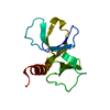
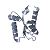
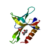
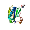
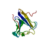
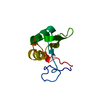
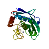
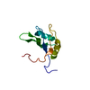
| Title | The crystal structure of villin domain 6 | ||||||
 Components Components | Villin-1 | ||||||
 Keywords Keywords | STRUCTURAL PROTEIN / actin binding protein / Villin head piece / villin / gelsolin / Actin capping / Actin-binding / Calcium / Cytoplasm / Cytoskeleton | ||||||
| Function / homology |  Function and homology informationterminal web assembly / intestinal D-glucose absorption / regulation of microvillus length / regulation of actin nucleation / cytoplasmic actin-based contraction involved in cell motility / positive regulation of actin filament depolymerization / lysophosphatidic acid binding / regulation of lamellipodium morphogenesis / filopodium tip / positive regulation of lamellipodium morphogenesis ...terminal web assembly / intestinal D-glucose absorption / regulation of microvillus length / regulation of actin nucleation / cytoplasmic actin-based contraction involved in cell motility / positive regulation of actin filament depolymerization / lysophosphatidic acid binding / regulation of lamellipodium morphogenesis / filopodium tip / positive regulation of lamellipodium morphogenesis / positive regulation of actin filament bundle assembly / actin filament severing / regulation of wound healing / actin filament capping / barbed-end actin filament capping / positive regulation of multicellular organism growth / actin polymerization or depolymerization / actin filament depolymerization / cellular response to hepatocyte growth factor stimulus / positive regulation of epithelial cell migration / actin filament bundle / microvillus / cysteine-type endopeptidase inhibitor activity involved in apoptotic process / brush border / cellular response to epidermal growth factor stimulus / ruffle / epithelial cell differentiation / phosphatidylinositol-4,5-bisphosphate binding / actin filament polymerization / filopodium / positive regulation of protein localization to plasma membrane / response to bacterium / epidermal growth factor receptor signaling pathway / actin filament binding / actin cytoskeleton / lamellipodium / regulation of cell shape / protein-containing complex assembly / positive regulation of cell migration / apoptotic process / calcium ion binding / protein homodimerization activity / extracellular exosome / identical protein binding / plasma membrane / cytoplasm Function and homology informationterminal web assembly / intestinal D-glucose absorption / regulation of microvillus length / regulation of actin nucleation / cytoplasmic actin-based contraction involved in cell motility / positive regulation of actin filament depolymerization / lysophosphatidic acid binding / regulation of lamellipodium morphogenesis / filopodium tip / positive regulation of lamellipodium morphogenesis ...terminal web assembly / intestinal D-glucose absorption / regulation of microvillus length / regulation of actin nucleation / cytoplasmic actin-based contraction involved in cell motility / positive regulation of actin filament depolymerization / lysophosphatidic acid binding / regulation of lamellipodium morphogenesis / filopodium tip / positive regulation of lamellipodium morphogenesis / positive regulation of actin filament bundle assembly / actin filament severing / regulation of wound healing / actin filament capping / barbed-end actin filament capping / positive regulation of multicellular organism growth / actin polymerization or depolymerization / actin filament depolymerization / cellular response to hepatocyte growth factor stimulus / positive regulation of epithelial cell migration / actin filament bundle / microvillus / cysteine-type endopeptidase inhibitor activity involved in apoptotic process / brush border / cellular response to epidermal growth factor stimulus / ruffle / epithelial cell differentiation / phosphatidylinositol-4,5-bisphosphate binding / actin filament polymerization / filopodium / positive regulation of protein localization to plasma membrane / response to bacterium / epidermal growth factor receptor signaling pathway / actin filament binding / actin cytoskeleton / lamellipodium / regulation of cell shape / protein-containing complex assembly / positive regulation of cell migration / apoptotic process / calcium ion binding / protein homodimerization activity / extracellular exosome / identical protein binding / plasma membrane / cytoplasmSimilarity search - Function | ||||||
| Biological species |  Homo sapiens (human) Homo sapiens (human) | ||||||
| Method | X-RAY DIFFRACTION / SYNCHROTRON / MOLECULAR REPLACEMENT / molecular replacement / Resolution: 2 Å | ||||||
 Authors Authors | Wang, H. / Burtnick, L.D. / Robinson, R.C. | ||||||
 Citation Citation | Journal: J.Biol.Chem. / Year: 2009 Title: Helix-straightening as an activation mechanism in the gelsolin superfamily of actin regulatory proteins Authors: Wang, H. / Chumnarnsilpa, S. / Loonchanta, A. / Li, Q. / Kuan, Y.M. / Robine, S. / Larsson, M. / Mihalek, I. / Burtnick, L.D. / Robinson, R.C. | ||||||
| History |
|
- Structure visualization
| Structure viewer | Molecule: MolmilJmol/JSmol |
|---|
- Downloads & links
-Download
| PDBx/mmCIF format | 3fg7.cif.gz | 114.5 KB | Display | PDBx/mmCIF format |
|---|---|---|---|---|
| PDB format | pdb3fg7.ent.gz | 82.4 KB | Display | PDB format |
| PDBx/mmJSON format | 3fg7.json.gz | Tree view | PDBx/mmJSON format | |
| Others |  Other downloads Other downloads |
-Validation report
| Arichive directory | https://data.pdbj.org/pub/pdb/validation_reports/fg/3fg7ftp://data.pdbj.org/pub/pdb/validation_reports/fg/3fg7 | HTTPS FTP |
|---|
-Related structure data
| Related structure data |  1p8xS S: Starting model for refinement |
|---|---|
| Similar structure data |


-Links
 PDBj
PDBj
- Assembly
| Deposited unit | 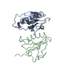
| |||||||||||||||||||||||||||||||||||||||||||||||||||||||||||||||||||||||||||||||||||||||||||||||||||||||||
|---|---|---|---|---|---|---|---|---|---|---|---|---|---|---|---|---|---|---|---|---|---|---|---|---|---|---|---|---|---|---|---|---|---|---|---|---|---|---|---|---|---|---|---|---|---|---|---|---|---|---|---|---|---|---|---|---|---|---|---|---|---|---|---|---|---|---|---|---|---|---|---|---|---|---|---|---|---|---|---|---|---|---|---|---|---|---|---|---|---|---|---|---|---|---|---|---|---|---|---|---|---|---|---|---|---|---|
| 1 | 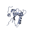
| |||||||||||||||||||||||||||||||||||||||||||||||||||||||||||||||||||||||||||||||||||||||||||||||||||||||||
| 2 | 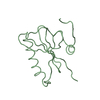
| |||||||||||||||||||||||||||||||||||||||||||||||||||||||||||||||||||||||||||||||||||||||||||||||||||||||||
| Unit cell |
| |||||||||||||||||||||||||||||||||||||||||||||||||||||||||||||||||||||||||||||||||||||||||||||||||||||||||
| Noncrystallographic symmetry (NCS) | NCS domain:
NCS domain segments:
NCS ensembles :
|
-Components
| #1: Protein | Mass: 44981.551 Da / Num. of mol.: 2 / Fragment: Gelsolin-like repeats 4, 5, and 6 Source method: isolated from a genetically manipulated source Source: (gene. exp.) Homo sapiens (human) / Gene: VIL, VIL1 / Plasmid: pSY5 / Production host:  Escherichia coli (E. coli) / References: UniProt: P09327 Escherichia coli (E. coli) / References: UniProt: P09327#2: Water | ChemComp-HOH / | Water Mass: 18.015 Da / Num. of mol.: 160 / Source method: isolated from a natural source / Formula: H2O Mass: 18.015 Da / Num. of mol.: 160 / Source method: isolated from a natural source / Formula: H2O |
|---|
-Experimental details
-Experiment
| Experiment | Method: X-RAY DIFFRACTION / Number of used crystals: 1 |
|---|
- Sample preparation
| Crystal | Density Matthews: 2.63 Å3/Da / Density % sol: 53.17 % |
|---|---|
| Crystal grow | Temperature: 277 K / Method: vapor diffusion, hanging drop / pH: 5 Details: The protein(10mg/ml)was mixed in a 1:1 ratio(v/v)with a reservoir solution containing 15%(w/v)PEG8000, 100mM NaOAc buffer, pH 5.0, VAPOR DIFFUSION, HANGING DROP, temperature 277KK |
-Data collection
| Diffraction | Mean temperature: 100 K | |||||||||||||||||||||||||||||||||||||||||||||||||||||||||||||||||||||||||||||
|---|---|---|---|---|---|---|---|---|---|---|---|---|---|---|---|---|---|---|---|---|---|---|---|---|---|---|---|---|---|---|---|---|---|---|---|---|---|---|---|---|---|---|---|---|---|---|---|---|---|---|---|---|---|---|---|---|---|---|---|---|---|---|---|---|---|---|---|---|---|---|---|---|---|---|---|---|---|---|
| Diffraction source | Source: SYNCHROTRON / Site: NSRRC  / Beamline: BL13B1 / Wavelength: 1 / Beamline: BL13B1 / Wavelength: 1 | |||||||||||||||||||||||||||||||||||||||||||||||||||||||||||||||||||||||||||||
| Detector | Type: ADSC QUANTUM 315 / Detector: CCD / Date: Mar 31, 2008 | |||||||||||||||||||||||||||||||||||||||||||||||||||||||||||||||||||||||||||||
| Radiation | Protocol: SINGLE WAVELENGTH / Monochromatic (M) / Laue (L): M / Scattering type: x-ray | |||||||||||||||||||||||||||||||||||||||||||||||||||||||||||||||||||||||||||||
| Radiation wavelength | Wavelength: 1 Å / Relative weight: 1 | |||||||||||||||||||||||||||||||||||||||||||||||||||||||||||||||||||||||||||||
| Reflection | Redundancy: 4.8 % / Av σ(I) over netI: 44.8 / Number: 80501 / Rmerge(I) obs: 0.048 / Χ2: 1.71 / D res high: 2 Å / D res low: 30 Å / Num. obs: 16865 / % possible obs: 99.6 | |||||||||||||||||||||||||||||||||||||||||||||||||||||||||||||||||||||||||||||
| Diffraction reflection shell |
| |||||||||||||||||||||||||||||||||||||||||||||||||||||||||||||||||||||||||||||
| Reflection | Resolution: 2→30 Å / Num. obs: 16865 / % possible obs: 99.6 % / Redundancy: 4.8 % / Rmerge(I) obs: 0.048 | |||||||||||||||||||||||||||||||||||||||||||||||||||||||||||||||||||||||||||||
| Reflection shell | Resolution: 2→2.07 Å / Redundancy: 3.5 % / Rmerge(I) obs: 0.282 / % possible all: 98.8 |
-Phasing
| Phasing | Method: molecular replacement | |||||||||
|---|---|---|---|---|---|---|---|---|---|---|
| Phasing MR | Model details: Phaser MODE: MR_AUTO
|
- Processing
| Software |
| ||||||||||||||||||||||||||||||||||||||||||||||||||||||||||||||||||||||||||||||||||||||||||||||||||||||||||||||||||||||||||||||||||||||||||||||||||||||||||||||||||||||||||
|---|---|---|---|---|---|---|---|---|---|---|---|---|---|---|---|---|---|---|---|---|---|---|---|---|---|---|---|---|---|---|---|---|---|---|---|---|---|---|---|---|---|---|---|---|---|---|---|---|---|---|---|---|---|---|---|---|---|---|---|---|---|---|---|---|---|---|---|---|---|---|---|---|---|---|---|---|---|---|---|---|---|---|---|---|---|---|---|---|---|---|---|---|---|---|---|---|---|---|---|---|---|---|---|---|---|---|---|---|---|---|---|---|---|---|---|---|---|---|---|---|---|---|---|---|---|---|---|---|---|---|---|---|---|---|---|---|---|---|---|---|---|---|---|---|---|---|---|---|---|---|---|---|---|---|---|---|---|---|---|---|---|---|---|---|---|---|---|---|---|---|---|
| Refinement | Method to determine structure: MOLECULAR REPLACEMENT Starting model: PDB entry 1P8X Resolution: 2→23.91 Å / Cor.coef. Fo:Fc: 0.956 / Cor.coef. Fo:Fc free: 0.92 / Occupancy max: 1 / Occupancy min: 1 / SU B: 9.303 / SU ML: 0.12 / Cross valid method: THROUGHOUT / ESU R Free: 0.173 / Stereochemistry target values: MAXIMUM LIKELIHOOD / Details: HYDROGENS HAVE BEEN ADDED IN THE RIDING POSITIONS
| ||||||||||||||||||||||||||||||||||||||||||||||||||||||||||||||||||||||||||||||||||||||||||||||||||||||||||||||||||||||||||||||||||||||||||||||||||||||||||||||||||||||||||
| Solvent computation | Ion probe radii: 0.8 Å / Shrinkage radii: 0.8 Å / VDW probe radii: 1.4 Å / Solvent model: MASK | ||||||||||||||||||||||||||||||||||||||||||||||||||||||||||||||||||||||||||||||||||||||||||||||||||||||||||||||||||||||||||||||||||||||||||||||||||||||||||||||||||||||||||
| Displacement parameters | Biso mean: 38.799 Å2
| ||||||||||||||||||||||||||||||||||||||||||||||||||||||||||||||||||||||||||||||||||||||||||||||||||||||||||||||||||||||||||||||||||||||||||||||||||||||||||||||||||||||||||
| Refinement step | Cycle: LAST / Resolution: 2→23.91 Å
| ||||||||||||||||||||||||||||||||||||||||||||||||||||||||||||||||||||||||||||||||||||||||||||||||||||||||||||||||||||||||||||||||||||||||||||||||||||||||||||||||||||||||||
| Refine LS restraints |
| ||||||||||||||||||||||||||||||||||||||||||||||||||||||||||||||||||||||||||||||||||||||||||||||||||||||||||||||||||||||||||||||||||||||||||||||||||||||||||||||||||||||||||
| Refine LS restraints NCS | Dom-ID: 1 / Auth asym-ID: B / Refine-ID: X-RAY DIFFRACTION
| ||||||||||||||||||||||||||||||||||||||||||||||||||||||||||||||||||||||||||||||||||||||||||||||||||||||||||||||||||||||||||||||||||||||||||||||||||||||||||||||||||||||||||
| LS refinement shell | Resolution: 2.003→2.055 Å / Total num. of bins used: 20
|
