 Movie
Movie Controller

[English] 日本語
 Yorodumi
- PDB-1nph: Gelsolin Domains 4-6 in Active, Actin Free Conformation Identifie... -
+ Open data

- Basic information
| Entry | Database: PDB / ID: 1nph | ||||||
|---|---|---|---|---|---|---|---|
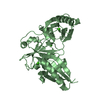
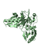
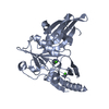
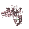
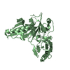
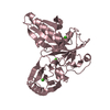
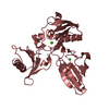
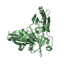
| Title | Gelsolin Domains 4-6 in Active, Actin Free Conformation Identifies Sites of Regulatory Calcium Ions | ||||||
 Components Components | Gelsolin | ||||||
 Keywords Keywords | PROTEIN BINDING / BETA SHEET | ||||||
| Function / homology |  Function and homology information Function and homology informationglial filament / apical ectoplasmic specialization / basal ectoplasmic specialization / Caspase-mediated cleavage of cytoskeletal proteins / striated muscle atrophy / regulation of establishment of T cell polarity / regulation of plasma membrane raft polarization / regulation of receptor clustering / renal protein absorption / positive regulation of keratinocyte apoptotic process ...glial filament / apical ectoplasmic specialization / basal ectoplasmic specialization / Caspase-mediated cleavage of cytoskeletal proteins / striated muscle atrophy / regulation of establishment of T cell polarity / regulation of plasma membrane raft polarization / regulation of receptor clustering / renal protein absorption / positive regulation of keratinocyte apoptotic process / positive regulation of protein processing in phagocytic vesicle / positive regulation of actin nucleation / phosphatidylinositol 3-kinase catalytic subunit binding / actin cap / sequestering of actin monomers / regulation of podosome assembly / myosin II binding / negative regulation of viral entry into host cell / actin filament severing / actin filament capping / barbed-end actin filament capping / actin polymerization or depolymerization / actin filament depolymerization / cell projection assembly / cardiac muscle cell contraction / podosome / positive regulation of p38MAPK cascade / relaxation of cardiac muscle / phagocytosis, engulfment / cortical actin cytoskeleton / positive regulation of cardiac muscle hypertrophy / hepatocyte apoptotic process / cilium assembly / sarcoplasm / phagocytic vesicle / vesicle-mediated transport / ruffle / cellular response to cadmium ion / phosphatidylinositol-4,5-bisphosphate binding / actin filament polymerization / response to muscle stretch / Neutrophil degranulation / central nervous system development / actin filament organization / protein destabilization / cellular response to type II interferon / actin filament binding / actin cytoskeleton / lamellipodium / myelin sheath / actin binding / actin cytoskeleton organization / amyloid fibril formation / calcium ion binding / positive regulation of gene expression / perinuclear region of cytoplasm / protein-containing complex / extracellular space / extracellular region / nucleus / plasma membrane / cytosol / cytoplasmSimilarity search - Function | ||||||
| Biological species |  Mus musculus (house mouse) Mus musculus (house mouse) | ||||||
| Method | X-RAY DIFFRACTION / SYNCHROTRON / MOLECULAR REPLACEMENT / Resolution: 3 Å | ||||||
 Authors Authors | Kolappan, S. / Gooch, J.T. / Weeds, A.G. / McLaughlin, P.J. | ||||||
 Citation Citation | Journal: J.Mol.Biol. / Year: 2003 Title: Gelsolin Domains 4-6 in Active, Actin-Free Conformation Identifies Sites of Regulatory Calcium Ions Authors: Kolappan, S. / Gooch, J.T. / Weeds, A.G. / McLaughlin, P.J. | ||||||
| History |
|
- Structure visualization
| Structure viewer | Molecule: MolmilJmol/JSmol |
|---|
- Downloads & links
-Download
| PDBx/mmCIF format | 1nph.cif.gz | 75.1 KB | Display | PDBx/mmCIF format |
|---|---|---|---|---|
| PDB format | pdb1nph.ent.gz | 54.8 KB | Display | PDB format |
| PDBx/mmJSON format | 1nph.json.gz | Tree view | PDBx/mmJSON format | |
| Others |  Other downloads Other downloads |
-Validation report
| Arichive directory | https://data.pdbj.org/pub/pdb/validation_reports/np/1nphftp://data.pdbj.org/pub/pdb/validation_reports/np/1nph | HTTPS FTP |
|---|
-Related structure data
| Related structure data |  1db0 S: Starting model for refinement |
|---|---|
| Similar structure data |

-Links
 PDBj
PDBj


- Assembly
| Deposited unit | 
| ||||||||
|---|---|---|---|---|---|---|---|---|---|
| 1 |
| ||||||||
| Unit cell |
|
-Components
| #1: Protein | / Actin-depolymerizing factor Mass: 36353.449 Da / Num. of mol.: 1 Source method: isolated from a genetically manipulated source Source: (gene. exp.) Mus musculus (house mouse) / Gene: GSN OR GSB / Plasmid: pET derivative pMW172 / Species (production host): Escherichia coli / Production host:  Escherichia coli BL21(DE3) (bacteria) / Strain (production host): BL21(DE3) / References: UniProt: P13020 Escherichia coli BL21(DE3) (bacteria) / Strain (production host): BL21(DE3) / References: UniProt: P13020 |
|---|---|
| #2: Chemical |   Mass: 40.078 Da / Num. of mol.: 2 / Source method: obtained synthetically / Formula: Ca Mass: 40.078 Da / Num. of mol.: 2 / Source method: obtained synthetically / Formula: Ca |
-Experimental details
-Experiment
| Experiment | Method: X-RAY DIFFRACTION / Number of used crystals: 1 |
|---|
- Sample preparation
| Crystal | Density Matthews: 4.52 Å3/Da / Density % sol: 72.56 % | |||||||||||||||||||||||||||||||||||
|---|---|---|---|---|---|---|---|---|---|---|---|---|---|---|---|---|---|---|---|---|---|---|---|---|---|---|---|---|---|---|---|---|---|---|---|---|
| Crystal grow | Temperature: 293 K / Method: vapor diffusion, hanging drop / pH: 8 Details: PEG 8000, calcium chloride, pH 8.0, VAPOR DIFFUSION, HANGING DROP, temperature 293.0K | |||||||||||||||||||||||||||||||||||
| Crystal grow | *PLUS Temperature: 20 ℃ | |||||||||||||||||||||||||||||||||||
| Components of the solutions | *PLUS
|
-Data collection
| Diffraction | Mean temperature: 100 K |
|---|---|
| Diffraction source | Source: SYNCHROTRON / Site: SRS  / Beamline: PX14.1 / Wavelength: 1.488 Å / Beamline: PX14.1 / Wavelength: 1.488 Å |
| Detector | Type: ADSC QUANTUM 4 / Detector: CCD / Date: Jan 31, 2002 / Details: mirror, Monochromator |
| Radiation | Monochromator: Liquid gallium cooled, bent, triangular, si 111 optimised for 1.488 (11.7 asymmetric cut) Protocol: SINGLE WAVELENGTH / Monochromatic (M) / Laue (L): M / Scattering type: x-ray |
| Radiation wavelength | Wavelength: 1.488 Å / Relative weight: 1 |
| Reflection | Resolution: 3→87.7 Å / Num. obs: 13119 / Observed criterion σ(I): 2 / Redundancy: 6.1 % / Rmerge(I) obs: 0.123 / Rsym value: 0.123 / Net I/σ(I): 5.2 |
| Reflection shell | Resolution: 3→3.16 Å / Rmerge(I) obs: 0.313 / Mean I/σ(I) obs: 2.3 / Rsym value: 0.313 |
| Reflection | *PLUS Num. obs: 13141 / % possible obs: 99.4 % / Rmerge(I) obs: 0.12 |
| Reflection shell | *PLUS Rmerge(I) obs: 0.31 |
- Processing
| Software |
| |||||||||||||||||||||||||||||||||||||||||||||||||||||||||||||||||||||||||||||
|---|---|---|---|---|---|---|---|---|---|---|---|---|---|---|---|---|---|---|---|---|---|---|---|---|---|---|---|---|---|---|---|---|---|---|---|---|---|---|---|---|---|---|---|---|---|---|---|---|---|---|---|---|---|---|---|---|---|---|---|---|---|---|---|---|---|---|---|---|---|---|---|---|---|---|---|---|---|---|
| Refinement | Method to determine structure: MOLECULAR REPLACEMENT Starting model: PDB ENTRY 1DB0 1db0 Resolution: 3→25 Å / Isotropic thermal model: Isotropic / Cross valid method: THROUGHOUT / σ(F): 0 / Stereochemistry target values: Engh & Huber
| |||||||||||||||||||||||||||||||||||||||||||||||||||||||||||||||||||||||||||||
| Refine analyze |
| |||||||||||||||||||||||||||||||||||||||||||||||||||||||||||||||||||||||||||||
| Refinement step | Cycle: LAST / Resolution: 3→25 Å
| |||||||||||||||||||||||||||||||||||||||||||||||||||||||||||||||||||||||||||||
| Refine LS restraints |
| |||||||||||||||||||||||||||||||||||||||||||||||||||||||||||||||||||||||||||||
| LS refinement shell | Refine-ID: X-RAY DIFFRACTION
| |||||||||||||||||||||||||||||||||||||||||||||||||||||||||||||||||||||||||||||
| Refinement | *PLUS Highest resolution: 3 Å / Lowest resolution: 25 Å / Rfactor Rfree: 0.298 / Rfactor Rwork: 0.2452 | |||||||||||||||||||||||||||||||||||||||||||||||||||||||||||||||||||||||||||||
| Solvent computation | *PLUS | |||||||||||||||||||||||||||||||||||||||||||||||||||||||||||||||||||||||||||||
| Displacement parameters | *PLUS | |||||||||||||||||||||||||||||||||||||||||||||||||||||||||||||||||||||||||||||
| Refine LS restraints | *PLUS Type: c_bond_d / Dev ideal: 0.01 | |||||||||||||||||||||||||||||||||||||||||||||||||||||||||||||||||||||||||||||
| LS refinement shell | *PLUS Rfactor Rfree: 0.43 |
