 Movie
Movie Controller

[English] 日本語
 Yorodumi
- PDB-4wci: Crystal structure of the 1st SH3 domain from human CD2AP (CMS) in... -
+ Open data

- Basic information
| Entry | Database: PDB / ID: 4wci | ||||||
|---|---|---|---|---|---|---|---|
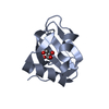
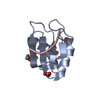
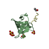
| Title | Crystal structure of the 1st SH3 domain from human CD2AP (CMS) in complex with a proline-rich peptide (aa 378-393) from human RIN3 | ||||||
 Components Components |
| ||||||
 Keywords Keywords |  SIGNALING PROTEIN / Endocytosis Adaptor protein Protein-peptide binary complex Kidney / Structural Genomics / Structural Genomics Consortium / SGC SIGNALING PROTEIN / Endocytosis Adaptor protein Protein-peptide binary complex Kidney / Structural Genomics / Structural Genomics Consortium / SGC | ||||||
| Function / homology |  Function and homology information Function and homology informationnegative regulation of mast cell chemotaxis / response to glial cell derived neurotrophic factor / negative regulation of small GTPase mediated signal transduction / transforming growth factor beta1 production / localization of cell / Rab protein signal transduction / negative regulation of transforming growth factor beta1 production / slit diaphragm / response to transforming growth factor beta / podocyte differentiation ...negative regulation of mast cell chemotaxis / response to glial cell derived neurotrophic factor / negative regulation of small GTPase mediated signal transduction / transforming growth factor beta1 production / localization of cell / Rab protein signal transduction / negative regulation of transforming growth factor beta1 production / slit diaphragm / response to transforming growth factor beta / podocyte differentiation / regulation of vesicle size / endothelium development / immunological synapse formation / nerve growth factor signaling pathway / cell-cell adhesion mediated by cadherin / collateral sprouting / protein heterooligomerization / renal albumin absorption / substrate-dependent cell migration, cell extension / membrane organization / phosphatidylinositol 3-kinase regulatory subunit binding / RAB GEFs exchange GTP for GDP on RABs / cell-cell junction organization / filopodium assembly / negative regulation of receptor internalization / podosome / Nephrin family interactions / clathrin binding / maintenance of blood-brain barrier / nuclear envelope lumen / glucose import / cell leading edge / filamentous actin / neurotrophin TRK receptor signaling pathway / centriolar satellite / protein secretion / endocytic vesicle / lymph node development / adipose tissue development / stress-activated MAPK cascade / ruffle / ERK1 and ERK2 cascade / actin filament polymerization / GTPase activator activity / guanyl-nucleotide exchange factor activity / trans-Golgi network membrane / liver development / phosphatidylinositol 3-kinase/protein kinase B signal transduction / actin filament organization / positive regulation of protein secretion / regulation of actin cytoskeleton organization / synapse organization / response to virus / regulation of synaptic plasticity / protein catabolic process / response to insulin / neuromuscular junction / lipid metabolic process / structural constituent of cytoskeleton / fibrillar center / small GTPase binding / SH3 domain binding / response to wounding / endocytosis / positive regulation of protein localization to nucleus / male gonad development / actin filament binding / cell migration / actin cytoskeleton / late endosome / T cell receptor signaling pathway / growth cone / cytoplasmic vesicle / protein-containing complex assembly / response to oxidative stress / vesicle / negative regulation of neuron apoptotic process / cell population proliferation / early endosome / cadherin binding / inflammatory response / cell cycle / axon / cell division / neuronal cell body / apoptotic process / dendrite / signal transduction / extracellular exosome / identical protein binding / plasma membrane / cytosol / cytoplasmSimilarity search - Function | ||||||
| Biological species |  Homo sapiens (human) Homo sapiens (human) | ||||||
| Method | X-RAY DIFFRACTION / SYNCHROTRON / MOLECULAR REPLACEMENT / Resolution: 1.65 Å | ||||||
 Authors Authors | Rouka, E. / Simister, P.C. / Janning, M. / Kirsch, K.H. / Krojer, T. / Knapp, S. / von Delft, F. / Arrowsmith, C.H. / Edwards, A.M. / Bountra, C. ...Rouka, E. / Simister, P.C. / Janning, M. / Kirsch, K.H. / Krojer, T. / Knapp, S. / von Delft, F. / Arrowsmith, C.H. / Edwards, A.M. / Bountra, C. / Feller, S.M. / Structural Genomics Consortium (SGC) | ||||||
 Citation Citation | Journal: J.Biol.Chem. / Year: 2015 Title: Differential Recognition Preferences of the Three Src Homology 3 (SH3) Domains from the Adaptor CD2-associated Protein (CD2AP) and Direct Association with Ras and Rab Interactor 3 (RIN3). Authors: Rouka, E. / Simister, P.C. / Janning, M. / Kumbrink, J. / Konstantinou, T. / Muniz, J.R. / Joshi, D. / O'Reilly, N. / Volkmer, R. / Ritter, B. / Knapp, S. / von Delft, F. / Kirsch, K.H. / Feller, S.M. | ||||||
| History |
|
- Structure visualization
| Structure viewer | Molecule: MolmilJmol/JSmol |
|---|
- Downloads & links
-Download
| PDBx/mmCIF format | 4wci.cif.gz | 109 KB | Display | PDBx/mmCIF format |
|---|---|---|---|---|
| PDB format | pdb4wci.ent.gz | 84.2 KB | Display | PDB format |
| PDBx/mmJSON format | 4wci.json.gz | Tree view | PDBx/mmJSON format | |
| Others |  Other downloads Other downloads |
-Validation report
| Arichive directory | https://data.pdbj.org/pub/pdb/validation_reports/wc/4wciftp://data.pdbj.org/pub/pdb/validation_reports/wc/4wci | HTTPS FTP |
|---|
-Related structure data
| Related structure data |  3u23C  2j6fS C: citing same article ( S: Starting model for refinement |
|---|---|
| Similar structure data |


-Links
 PDBj
PDBj
- Assembly
| Deposited unit | 
| ||||||||
|---|---|---|---|---|---|---|---|---|---|
| 1 | 
| ||||||||
| 2 | 
| ||||||||
| 3 | 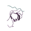
| ||||||||
| Unit cell |
| ||||||||
| Components on special symmetry positions |
|
-Components
| #1: Protein | Mass: 7593.522 Da / Num. of mol.: 3 / Fragment: UNP residues 1-60 Source method: isolated from a genetically manipulated source Source: (gene. exp.) Homo sapiens (human) / Gene: CD2AP / Plasmid: pGEX-6P-1 / Production host:  Escherichia coli BL21(DE3) (bacteria) / References: UniProt: Q9Y5K6 Escherichia coli BL21(DE3) (bacteria) / References: UniProt: Q9Y5K6#2: Protein/peptide | Mass: 1825.143 Da / Num. of mol.: 3 / Source method: obtained synthetically / Source: (synth.) Homo sapiens (human) / References: UniProt: Q8TB24#3: Chemical | Sulfate  Mass: 96.063 Da / Num. of mol.: 2 / Source method: obtained synthetically / Formula: SO4 Mass: 96.063 Da / Num. of mol.: 2 / Source method: obtained synthetically / Formula: SO4#4: Water | ChemComp-HOH / | Water Mass: 18.015 Da / Num. of mol.: 279 / Source method: isolated from a natural source / Formula: H2O Mass: 18.015 Da / Num. of mol.: 279 / Source method: isolated from a natural source / Formula: H2O |
|---|
-Experimental details
-Experiment
| Experiment | Method: X-RAY DIFFRACTION |
|---|
- Sample preparation
| Crystal | Density Matthews: 2.25 Å3/Da / Density % sol: 45.45 % |
|---|---|
| Crystal grow | Temperature: 293 K / Method: vapor diffusion, sitting drop Details: 0.1 M Ca(ac) pH 4.2, 0.2 M Li2SO4 and 25% PEG 10 000 |
-Data collection
| Diffraction | Mean temperature: 100 K |
|---|---|
| Diffraction source | Source: SYNCHROTRON / Site: Diamond  / Beamline: I04 / Wavelength: 0.9173 Å / Beamline: I04 / Wavelength: 0.9173 Å |
| Detector | Type: PSI PILATUS 6M / Detector: PIXEL / Date: Dec 20, 2011 |
| Radiation | Protocol: SINGLE WAVELENGTH / Monochromatic (M) / Laue (L): M / Scattering type: x-ray |
| Radiation wavelength | Wavelength: 0.9173 Å / Relative weight: 1 |
| Reflection | Resolution: 1.65→48.19 Å / Num. all: 29731 / Num. obs: 29622 / % possible obs: 97.7 % / Redundancy: 3.5 % / Rmerge(I) obs: 0.132 / Net I/σ(I): 6.6 |
| Reflection shell | Resolution: 1.65→1.74 Å / Redundancy: 3.5 % / Rmerge(I) obs: 0.479 / Mean I/σ(I) obs: 2.4 / % possible all: 93 |
- Processing
| Software |
| |||||||||||||||||||||||||||||||||||||||||||||||||||||||||||||||||||||||||||||||||||||||||||||||||||||||||||||||||||||||||||||||||||||||||||||||||||||||||||||||||||||||||||||||
|---|---|---|---|---|---|---|---|---|---|---|---|---|---|---|---|---|---|---|---|---|---|---|---|---|---|---|---|---|---|---|---|---|---|---|---|---|---|---|---|---|---|---|---|---|---|---|---|---|---|---|---|---|---|---|---|---|---|---|---|---|---|---|---|---|---|---|---|---|---|---|---|---|---|---|---|---|---|---|---|---|---|---|---|---|---|---|---|---|---|---|---|---|---|---|---|---|---|---|---|---|---|---|---|---|---|---|---|---|---|---|---|---|---|---|---|---|---|---|---|---|---|---|---|---|---|---|---|---|---|---|---|---|---|---|---|---|---|---|---|---|---|---|---|---|---|---|---|---|---|---|---|---|---|---|---|---|---|---|---|---|---|---|---|---|---|---|---|---|---|---|---|---|---|---|---|---|
| Refinement | Method to determine structure: MOLECULAR REPLACEMENT Starting model: 2J6F Resolution: 1.65→48.186 Å / SU ML: 0.2 / Cross valid method: FREE R-VALUE / σ(F): 1.36 / Phase error: 22.8 / Stereochemistry target values: ML
| |||||||||||||||||||||||||||||||||||||||||||||||||||||||||||||||||||||||||||||||||||||||||||||||||||||||||||||||||||||||||||||||||||||||||||||||||||||||||||||||||||||||||||||||
| Solvent computation | Shrinkage radii: 0.9 Å / VDW probe radii: 1.11 Å / Solvent model: FLAT BULK SOLVENT MODEL | |||||||||||||||||||||||||||||||||||||||||||||||||||||||||||||||||||||||||||||||||||||||||||||||||||||||||||||||||||||||||||||||||||||||||||||||||||||||||||||||||||||||||||||||
| Refinement step | Cycle: LAST / Resolution: 1.65→48.186 Å
| |||||||||||||||||||||||||||||||||||||||||||||||||||||||||||||||||||||||||||||||||||||||||||||||||||||||||||||||||||||||||||||||||||||||||||||||||||||||||||||||||||||||||||||||
| Refine LS restraints |
| |||||||||||||||||||||||||||||||||||||||||||||||||||||||||||||||||||||||||||||||||||||||||||||||||||||||||||||||||||||||||||||||||||||||||||||||||||||||||||||||||||||||||||||||
| LS refinement shell |
| |||||||||||||||||||||||||||||||||||||||||||||||||||||||||||||||||||||||||||||||||||||||||||||||||||||||||||||||||||||||||||||||||||||||||||||||||||||||||||||||||||||||||||||||
| Refinement TLS params. | Method: refined / Refine-ID: X-RAY DIFFRACTION
| |||||||||||||||||||||||||||||||||||||||||||||||||||||||||||||||||||||||||||||||||||||||||||||||||||||||||||||||||||||||||||||||||||||||||||||||||||||||||||||||||||||||||||||||
| Refinement TLS group |
|
