 Movie
Movie Controller

[English] 日本語
 Yorodumi
- PDB-2uzc: Structure of human PDLIM5 in complex with the C-terminal peptide ... -
+ Open data

- Basic information
| Entry | Database: PDB / ID: 2uzc | ||||||
|---|---|---|---|---|---|---|---|
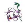
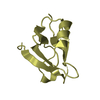
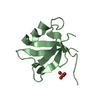
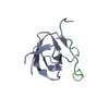
| Title | Structure of human PDLIM5 in complex with the C-terminal peptide of human alpha-actinin-1 | ||||||
 Components Components | PDZ AND LIM DOMAIN 5 | ||||||
 Keywords Keywords |  SIGNALING PROTEIN / METAL-BINDING / ENIGMA HOMOLOG / PHOSPHORYLATION / LIM DOMAIN / PDZ DOMAIN SIGNALING PROTEIN / METAL-BINDING / ENIGMA HOMOLOG / PHOSPHORYLATION / LIM DOMAIN / PDZ DOMAIN | ||||||
| Function / homology |  Function and homology information Function and homology informationmuscle structure development / cell growth involved in cardiac muscle cell development / : / muscle alpha-actinin binding / cadherin binding involved in cell-cell adhesion / regulation of dendritic spine morphogenesis / actinin binding / Neurexins and neuroligins / regulation of synapse assembly / filamentous actin ...muscle structure development / cell growth involved in cardiac muscle cell development / : / muscle alpha-actinin binding / cadherin binding involved in cell-cell adhesion / regulation of dendritic spine morphogenesis / actinin binding / Neurexins and neuroligins / regulation of synapse assembly / filamentous actin / stress fiber / protein kinase C binding / cell projection / adherens junction / Z disc / actin cytoskeleton / presynapse / heart development / actin binding / actin cytoskeleton organization / postsynaptic density / membrane / metal ion binding / cytosolSimilarity search - Function | ||||||
| Biological species |  HOMO SAPIENS (human) HOMO SAPIENS (human) | ||||||
| Method | X-RAY DIFFRACTION / SYNCHROTRON / MOLECULAR REPLACEMENT / Resolution: 1.5 Å | ||||||
 Authors Authors | Bunkoczi, G. / Elkins, J. / Salah, E. / Burgess-Brown, N. / Papagrigoriou, E. / Pike, A.C.W. / Turnbull, A. / Gileadi, O. / von Delft, F. / Arrowsmith, C.H. ...Bunkoczi, G. / Elkins, J. / Salah, E. / Burgess-Brown, N. / Papagrigoriou, E. / Pike, A.C.W. / Turnbull, A. / Gileadi, O. / von Delft, F. / Arrowsmith, C.H. / Edwards, A. / Sundstrom, M. / Weigelt, J. / Doyle, D. | ||||||
 Citation Citation | Journal: Protein Sci. / Year: 2010 Title: Unusual binding interactions in PDZ domain crystal structures help explain binding mechanisms. Authors: Elkins, J.M. / Gileadi, C. / Shrestha, L. / Phillips, C. / Wang, J. / Muniz, J.R. / Doyle, D.A. | ||||||
| History |
|
- Structure visualization
| Structure viewer | Molecule: MolmilJmol/JSmol |
|---|
- Downloads & links
-Download
| PDBx/mmCIF format | 2uzc.cif.gz | 106.1 KB | Display | PDBx/mmCIF format |
|---|---|---|---|---|
| PDB format | pdb2uzc.ent.gz | 82.6 KB | Display | PDB format |
| PDBx/mmJSON format | 2uzc.json.gz | Tree view | PDBx/mmJSON format | |
| Others |  Other downloads Other downloads |
-Validation report
| Arichive directory | https://data.pdbj.org/pub/pdb/validation_reports/uz/2uzcftp://data.pdbj.org/pub/pdb/validation_reports/uz/2uzc | HTTPS FTP |
|---|
-Related structure data
| Related structure data | 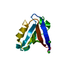 2pa1C 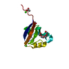 2pktSC  2pntC  2q3gC 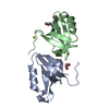 2v1wC  2w7rC S: Starting model for refinement C: citing same article ( |
|---|---|
| Similar structure data |


-Links
 PDBj
PDBj
- Assembly
| Deposited unit | 
| ||||||||||||||||||||
|---|---|---|---|---|---|---|---|---|---|---|---|---|---|---|---|---|---|---|---|---|---|
| 1 | 
| ||||||||||||||||||||
| 2 | 
| ||||||||||||||||||||
| 3 | 
| ||||||||||||||||||||
| 4 | 
| ||||||||||||||||||||
| 5 | 
| ||||||||||||||||||||
| Unit cell |
| ||||||||||||||||||||
| Noncrystallographic symmetry (NCS) | NCS oper:
|
-Components
| #1: Protein | Mass: 9317.584 Da / Num. of mol.: 5 / Fragment: RESIDUES 1-83 Source method: isolated from a genetically manipulated source Source: (gene. exp.) HOMO SAPIENS (human) / Plasmid: PNIC-BSA4 / Production host:  Escherichia coli BL21(DE3) (bacteria) / Variant (production host): R3 / References: UniProt: Q8WVK0, UniProt: Q96HC4*PLUS Escherichia coli BL21(DE3) (bacteria) / Variant (production host): R3 / References: UniProt: Q8WVK0, UniProt: Q96HC4*PLUS#2: Chemical | ChemComp-CL / Chloride  Mass: 35.453 Da / Num. of mol.: 6 / Source method: obtained synthetically / Formula: Cl Mass: 35.453 Da / Num. of mol.: 6 / Source method: obtained synthetically / Formula: Cl#3: Chemical | Ethylene glycol  Mass: 62.068 Da / Num. of mol.: 2 / Source method: obtained synthetically / Formula: C2H6O2 Mass: 62.068 Da / Num. of mol.: 2 / Source method: obtained synthetically / Formula: C2H6O2#4: Water | ChemComp-HOH / | Water Mass: 18.015 Da / Num. of mol.: 460 / Source method: isolated from a natural source / Formula: H2O Mass: 18.015 Da / Num. of mol.: 460 / Source method: isolated from a natural source / Formula: H2OSequence details | THE LAST 4 RESIDUES WERE TAGGED TO THE C-TERMINUS TO PROMOTE CRYSTAL CONTACTS | |
|---|
-Experimental details
-Experiment
| Experiment | Method: X-RAY DIFFRACTION / Number of used crystals: 1 |
|---|
- Sample preparation
| Crystal | Density Matthews: 2 Å3/Da / Density % sol: 38 % |
|---|---|
| Crystal grow | Details: 20% PEG3350 0.20 M KSCN |
-Data collection
| Diffraction | Mean temperature: 100 K |
|---|---|
| Diffraction source | Source: SYNCHROTRON / Site: SLS  / Beamline: X10SA / Wavelength: 0.9537 / Beamline: X10SA / Wavelength: 0.9537 |
| Detector | Type: MARRESEARCH / Detector: CCD / Date: Mar 3, 2007 / Details: MIRRORS |
| Radiation | Monochromator: SI111 / Protocol: SINGLE WAVELENGTH / Monochromatic (M) / Laue (L): M / Scattering type: x-ray |
| Radiation wavelength | Wavelength: 0.9537 Å / Relative weight: 1 |
| Reflection | Resolution: 1.5→46.4 Å / Num. obs: 49917 / % possible obs: 79.7 % / Observed criterion σ(I): 0 / Redundancy: 2.69 % / Rmerge(I) obs: 0.04 / Net I/σ(I): 17.2 |
| Reflection shell | Resolution: 1.5→1.6 Å / Redundancy: 0.83 % / Rmerge(I) obs: 0.38 / Mean I/σ(I) obs: 1.98 / % possible all: 37 |
- Processing
| Software |
| ||||||||||||||||||||||||||||||||||||||||||||||||||||||||||||||||||||||||||||||||||||||||||||||||||||||||||||||||||||||||||||||||||||||||||||||||||||||||||||||||||||||||||||||||||||||
|---|---|---|---|---|---|---|---|---|---|---|---|---|---|---|---|---|---|---|---|---|---|---|---|---|---|---|---|---|---|---|---|---|---|---|---|---|---|---|---|---|---|---|---|---|---|---|---|---|---|---|---|---|---|---|---|---|---|---|---|---|---|---|---|---|---|---|---|---|---|---|---|---|---|---|---|---|---|---|---|---|---|---|---|---|---|---|---|---|---|---|---|---|---|---|---|---|---|---|---|---|---|---|---|---|---|---|---|---|---|---|---|---|---|---|---|---|---|---|---|---|---|---|---|---|---|---|---|---|---|---|---|---|---|---|---|---|---|---|---|---|---|---|---|---|---|---|---|---|---|---|---|---|---|---|---|---|---|---|---|---|---|---|---|---|---|---|---|---|---|---|---|---|---|---|---|---|---|---|---|---|---|---|---|
| Refinement | Method to determine structure: MOLECULAR REPLACEMENT Starting model: PDB ENTRY 2PKT Resolution: 1.5→50 Å / Cor.coef. Fo:Fc: 0.97 / Cor.coef. Fo:Fc free: 0.952 / SU B: 3.312 / SU ML: 0.062 / TLS residual ADP flag: LIKELY RESIDUAL / Cross valid method: THROUGHOUT / ESU R: 0.092 / ESU R Free: 0.1 / Stereochemistry target values: MAXIMUM LIKELIHOOD / Details: HYDROGENS HAVE BEEN ADDED IN THE RIDING POSITIONS.
| ||||||||||||||||||||||||||||||||||||||||||||||||||||||||||||||||||||||||||||||||||||||||||||||||||||||||||||||||||||||||||||||||||||||||||||||||||||||||||||||||||||||||||||||||||||||
| Solvent computation | Ion probe radii: 0.8 Å / Shrinkage radii: 0.8 Å / VDW probe radii: 1.2 Å / Solvent model: MASK | ||||||||||||||||||||||||||||||||||||||||||||||||||||||||||||||||||||||||||||||||||||||||||||||||||||||||||||||||||||||||||||||||||||||||||||||||||||||||||||||||||||||||||||||||||||||
| Displacement parameters | Biso mean: 16.84 Å2
| ||||||||||||||||||||||||||||||||||||||||||||||||||||||||||||||||||||||||||||||||||||||||||||||||||||||||||||||||||||||||||||||||||||||||||||||||||||||||||||||||||||||||||||||||||||||
| Refinement step | Cycle: LAST / Resolution: 1.5→50 Å
| ||||||||||||||||||||||||||||||||||||||||||||||||||||||||||||||||||||||||||||||||||||||||||||||||||||||||||||||||||||||||||||||||||||||||||||||||||||||||||||||||||||||||||||||||||||||
| Refine LS restraints |
|