 Movie
Movie Controller

[English] 日本語
 Yorodumi
- PDB-6qjd: Crystal Structure of the truncated form of the third PDZ domain o... -
+ Open data

- Basic information
| Entry | Database: PDB / ID: 6qjd | |||||||||
|---|---|---|---|---|---|---|---|---|---|---|
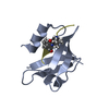
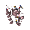
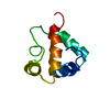
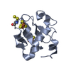
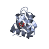
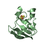
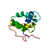
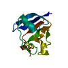
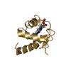
| Title | Crystal Structure of the truncated form of the third PDZ domain of PSD-95: residues 302-392 | |||||||||
 Components Components | Disks large homolog 4 | |||||||||
 Keywords Keywords |  SIGNALING PROTEIN / pdz domain SIGNALING PROTEIN / pdz domain | |||||||||
| Function / homology |  Function and homology information Function and homology informationLGI-ADAM interactions / P2Y1 nucleotide receptor binding / beta-1 adrenergic receptor binding / neuroligin family protein binding / NrCAM interactions / receptor localization to synapse / positive regulation of neuron projection arborization / regulation of grooming behavior / synaptic vesicle maturation / cerebellar mossy fiber ...LGI-ADAM interactions / P2Y1 nucleotide receptor binding / beta-1 adrenergic receptor binding / neuroligin family protein binding / NrCAM interactions / receptor localization to synapse / positive regulation of neuron projection arborization / regulation of grooming behavior / synaptic vesicle maturation / cerebellar mossy fiber / Synaptic adhesion-like molecules / cellular response to potassium ion / protein localization to synapse / vocalization behavior / neuron spine / AMPA glutamate receptor clustering / juxtaparanode region of axon / establishment or maintenance of epithelial cell apical/basal polarity / Trafficking of AMPA receptors / dendritic spine morphogenesis / negative regulation of receptor internalization / postsynaptic neurotransmitter receptor diffusion trapping / neuron projection terminus / RHO GTPases activate CIT / Assembly and cell surface presentation of NMDA receptors / acetylcholine receptor binding / Neurexins and neuroligins / neurotransmitter receptor localization to postsynaptic specialization membrane / Activation of Ca-permeable Kainate Receptor / cortical cytoskeleton / Signaling by ERBB4 / Negative regulation of NMDA receptor-mediated neuronal transmission / Unblocking of NMDA receptors, glutamate binding and activation / locomotory exploration behavior / regulation of NMDA receptor activity / social behavior / positive regulation of excitatory postsynaptic potential / Long-term potentiation / AMPA glutamate receptor complex / neuromuscular process controlling balance / excitatory synapse / D1 dopamine receptor binding / positive regulation of protein tyrosine kinase activity / positive regulation of synaptic transmission / ionotropic glutamate receptor binding / extrinsic component of cytoplasmic side of plasma membrane / Ras activation upon Ca2+ influx through NMDA receptor / dendrite cytoplasm / synaptic membrane / learning / PDZ domain binding / postsynaptic density membrane / adherens junction / regulation of long-term neuronal synaptic plasticity / neuromuscular junction / establishment of protein localization / cell-cell adhesion / kinase binding / endocytic vesicle membrane / synaptic vesicle / cell junction / nervous system development / positive regulation of cytosolic calcium ion concentration / chemical synaptic transmission / RAF/MAP kinase cascade / scaffold protein binding / postsynaptic membrane / basolateral plasma membrane / protein phosphatase binding / protein-containing complex assembly / dendritic spine / postsynaptic density / neuron projection / synapse / glutamatergic synapse / protein-containing complex binding / endoplasmic reticulum / signal transduction / plasma membrane / cytosol / cytoplasmSimilarity search - Function | |||||||||
| Biological species |  Homo sapiens (human) Homo sapiens (human) | |||||||||
| Method | X-RAY DIFFRACTION / SYNCHROTRON / MOLECULAR REPLACEMENT / molecular replacement / Resolution: 1.551 Å | |||||||||
 Authors Authors | Camara-Artigas, A. | |||||||||
| Funding support |  Spain, 1items Spain, 1items
| |||||||||
 Citation Citation | Journal: Acta Crystallogr D Struct Biol / Year: 2019 Title: Conformational changes in the third PDZ domain of the neuronal postsynaptic density protein 95. Authors: Camara-Artigas, A. / Murciano-Calles, J. / Martinez, J.C. | |||||||||
| History |
|
- Structure visualization
| Structure viewer | Molecule: MolmilJmol/JSmol |
|---|
- Downloads & links
-Download
| PDBx/mmCIF format | 6qjd.cif.gz | 193.9 KB | Display | PDBx/mmCIF format |
|---|---|---|---|---|
| PDB format | pdb6qjd.ent.gz | 158.8 KB | Display | PDB format |
| PDBx/mmJSON format | 6qjd.json.gz | Tree view | PDBx/mmJSON format | |
| Others |  Other downloads Other downloads |
-Validation report
| Arichive directory | https://data.pdbj.org/pub/pdb/validation_reports/qj/6qjdftp://data.pdbj.org/pub/pdb/validation_reports/qj/6qjd | HTTPS FTP |
|---|
-Related structure data
| Related structure data |  6qjfC 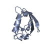 6qjgC 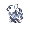 6qjiC 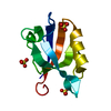 6qjjC  6qjkC  6qjlC 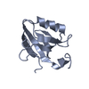 6qjnC 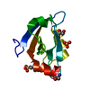 3k82S S: Starting model for refinement C: citing same article ( |
|---|---|
| Similar structure data |

-Links
 PDBj
PDBj


- Assembly
| Deposited unit | 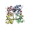
| ||||||||
|---|---|---|---|---|---|---|---|---|---|
| 1 | 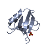
| ||||||||
| 2 | 
| ||||||||
| 3 | 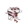
| ||||||||
| 4 | 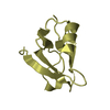
| ||||||||
| Unit cell |
|
-Components
| #1: Protein | Mass: 9909.155 Da / Num. of mol.: 4 / Fragment: PDZ domain Source method: isolated from a genetically manipulated source Source: (gene. exp.) Homo sapiens (human) / Gene: DLG4, PSD95 / Plasmid: pBAT4 / Production host:  Escherichia coli BL21(DE3) (bacteria) / References: UniProt: P78352 Escherichia coli BL21(DE3) (bacteria) / References: UniProt: P78352#2: Chemical | ChemComp-SO4 / Sulfate  Mass: 96.063 Da / Num. of mol.: 7 / Source method: obtained synthetically / Formula: SO4 Mass: 96.063 Da / Num. of mol.: 7 / Source method: obtained synthetically / Formula: SO4#3: Water | ChemComp-HOH / | Water Mass: 18.015 Da / Num. of mol.: 249 / Source method: isolated from a natural source / Formula: H2O Mass: 18.015 Da / Num. of mol.: 249 / Source method: isolated from a natural source / Formula: H2O |
|---|
-Experimental details
-Experiment
| Experiment | Method: X-RAY DIFFRACTION / Number of used crystals: 1 |
|---|
- Sample preparation
| Crystal | Density Matthews: 2.3 Å3/Da / Density % sol: 46.59 % / Mosaicity: 0.25 ° |
|---|---|
| Crystal grow | Temperature: 298 K / Method: vapor diffusion / pH: 6 / Details: 0.2 M ammonium sulphate, 30% PEG 4000 |
-Data collection
| Diffraction | Mean temperature: 100 K / Serial crystal experiment: N | |||||||||||||||||||||
|---|---|---|---|---|---|---|---|---|---|---|---|---|---|---|---|---|---|---|---|---|---|---|
| Diffraction source | Source: SYNCHROTRON / Site: ESRF  / Beamline: ID29 / Wavelength: 0.96864 Å / Beamline: ID29 / Wavelength: 0.96864 Å | |||||||||||||||||||||
| Detector | Type: DECTRIS PILATUS 6M / Detector: PIXEL / Date: Feb 16, 2014 / Details: Shutterless data collection | |||||||||||||||||||||
| Radiation | Monochromator: liquid nitrogen cooled channel-cut silicon monochromator Protocol: SINGLE WAVELENGTH / Monochromatic (M) / Laue (L): M / Scattering type: x-ray | |||||||||||||||||||||
| Radiation wavelength | Wavelength: 0.96864 Å / Relative weight: 1 | |||||||||||||||||||||
| Reflection | Resolution: 1.55→19.6 Å / Num. obs: 48816 / % possible obs: 94.1 % / Redundancy: 3.4 % / CC1/2: 0.983 / Rmerge(I) obs: 0.144 / Net I/σ(I): 5.4 | |||||||||||||||||||||
| Reflection shell |
|
-Phasing
| Phasing | Method: molecular replacement |
|---|
- Processing
| Software |
| ||||||||||||||||||||||||||||||||||||||||||||||||||||||||||||||||||||||||||||||||||||||||||||||||||||||||||||||||||||||||||||||
|---|---|---|---|---|---|---|---|---|---|---|---|---|---|---|---|---|---|---|---|---|---|---|---|---|---|---|---|---|---|---|---|---|---|---|---|---|---|---|---|---|---|---|---|---|---|---|---|---|---|---|---|---|---|---|---|---|---|---|---|---|---|---|---|---|---|---|---|---|---|---|---|---|---|---|---|---|---|---|---|---|---|---|---|---|---|---|---|---|---|---|---|---|---|---|---|---|---|---|---|---|---|---|---|---|---|---|---|---|---|---|---|---|---|---|---|---|---|---|---|---|---|---|---|---|---|---|---|
| Refinement | Method to determine structure: MOLECULAR REPLACEMENT Starting model: 3K82 Resolution: 1.551→19.6 Å / SU ML: 0.19 / Cross valid method: THROUGHOUT / σ(F): 1.01 / Phase error: 26.57
| ||||||||||||||||||||||||||||||||||||||||||||||||||||||||||||||||||||||||||||||||||||||||||||||||||||||||||||||||||||||||||||||
| Solvent computation | Shrinkage radii: 0.9 Å / VDW probe radii: 1.11 Å | ||||||||||||||||||||||||||||||||||||||||||||||||||||||||||||||||||||||||||||||||||||||||||||||||||||||||||||||||||||||||||||||
| Displacement parameters | Biso max: 73.89 Å2 / Biso mean: 17.1506 Å2 / Biso min: 4.05 Å2 | ||||||||||||||||||||||||||||||||||||||||||||||||||||||||||||||||||||||||||||||||||||||||||||||||||||||||||||||||||||||||||||||
| Refinement step | Cycle: final / Resolution: 1.551→19.6 Å
| ||||||||||||||||||||||||||||||||||||||||||||||||||||||||||||||||||||||||||||||||||||||||||||||||||||||||||||||||||||||||||||||
| LS refinement shell | Refine-ID: X-RAY DIFFRACTION / Rfactor Rfree error: 0 / Total num. of bins used: 17
|
