 Movie
Movie Controller

[English] 日本語
 Yorodumi
- PDB-4uyq: High resolution structure of the third cohesin ScaC in complex wi... -
+ Open data

- Basic information
| Entry | Database: PDB / ID: 4uyq | ||||||
|---|---|---|---|---|---|---|---|
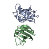
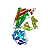
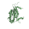
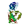
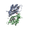
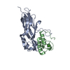
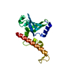
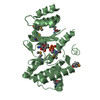
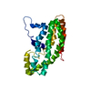
| Title | High resolution structure of the third cohesin ScaC in complex with the ScaB dockerin with a mutation in the C-terminal helix (IN to SI) from Acetivibrio cellulolyticus displaying a type I interaction. | ||||||
 Components Components |
| ||||||
 Keywords Keywords | CELL ADHESION/PROTEIN BINDING / CELL ADHESION-PROTEIN BINDING COMPLEX /  CELLULOSOME / TYPE 1 COHESIN-DOCKERIN INTEREACTIONS / ADAPTOR SCAFFOLDIN SCAB / ANCHORING SCAFFOLDING SCAC CELLULOSOME / TYPE 1 COHESIN-DOCKERIN INTEREACTIONS / ADAPTOR SCAFFOLDIN SCAB / ANCHORING SCAFFOLDING SCAC | ||||||
| Function / homology |  Function and homology information Function and homology informationpolysaccharide catabolic process / hydrolase activity, hydrolyzing O-glycosyl compounds / carbohydrate binding / extracellular region / metal ion bindingSimilarity search - Function | ||||||
| Biological species |  Acetivibrio cellulolyticus (bacteria) Acetivibrio cellulolyticus (bacteria) | ||||||
| Method | X-RAY DIFFRACTION / SYNCHROTRON / MOLECULAR REPLACEMENT / Resolution: 1.81 Å | ||||||
 Authors Authors | Cameron, K. / Fontes, C.M.G.A. / Najmudin, S. | ||||||
 Citation Citation | Journal: J. Biol. Chem. / Year: 2015 Title: Cell-surface Attachment of Bacterial Multienzyme Complexes Involves Highly Dynamic Protein-Protein Anchors. Authors: Cameron, K. / Najmudin, S. / Alves, V.D. / Bayer, E.A. / Smith, S.P. / Bule, P. / Waller, H. / Ferreira, L.M. / Gilbert, H.J. / Fontes, C.M. #1: Journal: Acta Crystallogr.,Sect.F / Year: 2012 Title: Purification, Crystallization and Preliminary X-Ray Characterization of the Acetivibrio Cellulolyticus Type I Cohesin Scac in Complex with the Scab Dockerin. Authors: Cameron, K. / Alves, V.D. / Bule, P. / Ferreira, L.M.A. / Fontes, C.M.G.A. / Najmudin, S. | ||||||
| History |
|
- Structure visualization
| Structure viewer | Molecule: MolmilJmol/JSmol |
|---|
- Downloads & links
-Download
| PDBx/mmCIF format | 4uyq.cif.gz | 105 KB | Display | PDBx/mmCIF format |
|---|---|---|---|---|
| PDB format | pdb4uyq.ent.gz | 81.1 KB | Display | PDB format |
| PDBx/mmJSON format | 4uyq.json.gz | Tree view | PDBx/mmJSON format | |
| Others |  Other downloads Other downloads |
-Validation report
| Arichive directory | https://data.pdbj.org/pub/pdb/validation_reports/uy/4uyqftp://data.pdbj.org/pub/pdb/validation_reports/uy/4uyq | HTTPS FTP |
|---|
-Related structure data
| Related structure data | 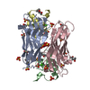 4uypSC  4uzp S: Starting model for refinement C: citing same article ( |
|---|---|
| Similar structure data |


-Links
 PDBj
PDBj
- Assembly
| Deposited unit | 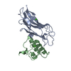
| ||||||||
|---|---|---|---|---|---|---|---|---|---|
| 1 |
| ||||||||
| Unit cell |
|
-Components
| #1: Protein | Mass: 16512.172 Da / Num. of mol.: 1 / Fragment: COHESIN, DOCKERIN, UNP RESIDUES 326-467 Source method: isolated from a genetically manipulated source Source: (gene. exp.) Acetivibrio cellulolyticus (bacteria) / Gene: scaC / Production host: Escherichia coli BL21(DE3) (bacteria) / Variant (production host): TUNER / References: UniProt: Q7WYN2 | ||||||
|---|---|---|---|---|---|---|---|
| #2: Protein | Mass: 8382.468 Da / Num. of mol.: 1 / Fragment: COHESIN, DOCKERIN, UNP RESIDUES 868-942 / Mutation: YES Source method: isolated from a genetically manipulated source Source: (gene. exp.) Acetivibrio cellulolyticus (bacteria) / Gene: scaB / Production host: Escherichia coli BL21(DE3) (bacteria) / Variant (production host): TUNER / References: UniProt: Q7WYN3 | ||||||
| #3: Chemical |   Mass: 40.078 Da / Num. of mol.: 3 / Source method: obtained synthetically / Formula: Ca Mass: 40.078 Da / Num. of mol.: 3 / Source method: obtained synthetically / Formula: Ca#4: Water | ChemComp-HOH / | Water Mass: 18.015 Da / Num. of mol.: 319 / Source method: isolated from a natural source / Formula: H2O Mass: 18.015 Da / Num. of mol.: 319 / Source method: isolated from a natural source / Formula: H2ONonpolymer details | CALCIUM ION (CA): CALCIUM IS FROM THE STORAGE BUFFER. | Sequence details | THE THIRD COHESIN CORRESPONDS TO RESIDUES 326 TO 427 OF THE ANCHORING SCAFFOLDIN SCAC. IT HAS A HIS ...THE THIRD COHESIN CORRESPOND | |
-Experimental details
-Experiment
| Experiment | Method: X-RAY DIFFRACTION / Number of used crystals: 1 |
|---|
- Sample preparation
| Crystal | Density Matthews: 2.68 Å3/Da / Density % sol: 54 % / Description: NONE |
|---|---|
| Crystal grow | pH: 7.5 Details: 0.2 M SODIUM ACETATE 0.1 M HEPES, PH 7.5 1.5 M K2HPO4 1.5 M NAH2PO4 |
-Data collection
| Diffraction | Mean temperature: 100 K |
|---|---|
| Diffraction source | Source: SYNCHROTRON / Site: Diamond  / Beamline: I03 / Wavelength: 0.9762 / Beamline: I03 / Wavelength: 0.9762 |
| Detector | Type: DECTRIS PILATUS 6M / Detector: PIXEL / Date: Jan 27, 2013 |
| Radiation | Protocol: SINGLE WAVELENGTH / Monochromatic (M) / Laue (L): M / Scattering type: x-ray |
| Radiation wavelength | Wavelength: 0.9762 Å / Relative weight: 1 |
| Reflection | Resolution: 1.81→68.17 Å / Num. obs: 23100 / % possible obs: 96.9 % / Observed criterion σ(I): 0 / Redundancy: 2.3 % / Rmerge(I) obs: 0.11 / Net I/σ(I): 5.35 |
| Reflection shell | Resolution: 1.81→1.88 Å / Redundancy: 2 % / Rmerge(I) obs: 0.42 / Mean I/σ(I) obs: 2.01 / % possible all: 93 |
- Processing
| Software |
| ||||||||||||||||||||||||||||||||||||||||||||||||||||||||||||||||||||||||||||||||||||||||||||||||||||||||||||||||||||||||||||||||||||||||||||||||||||||||||||||||||||||||||||||||||||||
|---|---|---|---|---|---|---|---|---|---|---|---|---|---|---|---|---|---|---|---|---|---|---|---|---|---|---|---|---|---|---|---|---|---|---|---|---|---|---|---|---|---|---|---|---|---|---|---|---|---|---|---|---|---|---|---|---|---|---|---|---|---|---|---|---|---|---|---|---|---|---|---|---|---|---|---|---|---|---|---|---|---|---|---|---|---|---|---|---|---|---|---|---|---|---|---|---|---|---|---|---|---|---|---|---|---|---|---|---|---|---|---|---|---|---|---|---|---|---|---|---|---|---|---|---|---|---|---|---|---|---|---|---|---|---|---|---|---|---|---|---|---|---|---|---|---|---|---|---|---|---|---|---|---|---|---|---|---|---|---|---|---|---|---|---|---|---|---|---|---|---|---|---|---|---|---|---|---|---|---|---|---|---|---|
| Refinement | Method to determine structure: MOLECULAR REPLACEMENT Starting model: PDB ENTRY 4UYP Resolution: 1.81→68.17 Å / Cor.coef. Fo:Fc: 0.969 / Cor.coef. Fo:Fc free: 0.944 / SU B: 5.239 / SU ML: 0.082 / Cross valid method: THROUGHOUT / ESU R: 0.106 / ESU R Free: 0.11 / Stereochemistry target values: MAXIMUM LIKELIHOOD Details: HYDROGENS HAVE BEEN ADDED IN THE RIDING POSITIONS. U VALUES WITH TLS ADDED. HIGH FLEXIBILITY OF THE N-TERMINUS LYSINE B 1 LEADING TO MULTIPLE CONFORMATIONS (THOUGH ONLY 2 ALTERNATE ONES ARE ...Details: HYDROGENS HAVE BEEN ADDED IN THE RIDING POSITIONS. U VALUES WITH TLS ADDED. HIGH FLEXIBILITY OF THE N-TERMINUS LYSINE B 1 LEADING TO MULTIPLE CONFORMATIONS (THOUGH ONLY 2 ALTERNATE ONES ARE GIVEN IN THIS PDB), LEADING TO REFINEMENT OF THE LYS IN THE THE AVERAGE POSITION.
| ||||||||||||||||||||||||||||||||||||||||||||||||||||||||||||||||||||||||||||||||||||||||||||||||||||||||||||||||||||||||||||||||||||||||||||||||||||||||||||||||||||||||||||||||||||||
| Solvent computation | Ion probe radii: 0.8 Å / Shrinkage radii: 0.8 Å / VDW probe radii: 1.2 Å / Solvent model: MASK | ||||||||||||||||||||||||||||||||||||||||||||||||||||||||||||||||||||||||||||||||||||||||||||||||||||||||||||||||||||||||||||||||||||||||||||||||||||||||||||||||||||||||||||||||||||||
| Displacement parameters | Biso mean: 23.735 Å2
| ||||||||||||||||||||||||||||||||||||||||||||||||||||||||||||||||||||||||||||||||||||||||||||||||||||||||||||||||||||||||||||||||||||||||||||||||||||||||||||||||||||||||||||||||||||||
| Refinement step | Cycle: LAST / Resolution: 1.81→68.17 Å
| ||||||||||||||||||||||||||||||||||||||||||||||||||||||||||||||||||||||||||||||||||||||||||||||||||||||||||||||||||||||||||||||||||||||||||||||||||||||||||||||||||||||||||||||||||||||
| Refine LS restraints |
|