 Movie
Movie Controller

[English] 日本語
 Yorodumi
- PDB-4uz8: The SeMet structure of the family 46 carbohydrate-binding module ... -
+ Open data

- Basic information
| Entry | Database: PDB / ID: 4uz8 | ||||||
|---|---|---|---|---|---|---|---|
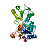
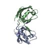
| Title | The SeMet structure of the family 46 carbohydrate-binding module (CBM46) of endo-beta-1,4-glucanase B (Cel5B) from Bacillus halodurans | ||||||
 Components Components | ENDO-BETA-1,4-GLUCANASE (CELULASE B) | ||||||
 Keywords Keywords | SUGAR BINDING PROTEIN / CARBOHYDRATE BINDING PROTEIN / CARBOHYDRATE-BINDING MODULE FAMILY 46 / CBM46 / CEL5B /  BACILLUS HALODURANS / SEMET DERIVATIVE BACILLUS HALODURANS / SEMET DERIVATIVE | ||||||
| Function / homology |  Function and homology information Function and homology informationglucan catabolic process / beta-glucosidase activity / cell surface / extracellular region / metal ion bindingSimilarity search - Function | ||||||
| Biological species |  BACILLUS HALODURANS (bacteria) BACILLUS HALODURANS (bacteria) | ||||||
| Method | X-RAY DIFFRACTION / SYNCHROTRON / SAD / Resolution: 2.3 Å | ||||||
 Authors Authors | Venditto, I. / Santos, H. / Ferreira, L.M.A. / Sakka, K. / Fontes, C.M.G.A. / Najmudin, S. | ||||||
 Citation Citation | Journal: J.Biol.Chem. / Year: 2015 Title: Family 46 Carbohydrate-Binding Modules Contribute to the Enzymatic Hydrolysis of Xyloglucan and Beta-1,3-1,4-Glucans Through Distinct Mechanisms. Authors: Venditto, I. / Najmudin, S. / Luis, A.S. / Ferreira, L.M. / Sakka, K. / Knox, J.P. / Gilbert, H.J. / Fontes, C.M. #1: Journal: Acta Crystallogr.,Sect.F / Year: 2014 Title: Overproduction, Purification, Crystallization and Preliminary X-Ray Characterization of the Family 46 Carbohydrate-Binding Module (Cbm46) of Endo-Beta-1,4-Glucanase B (Celb) from Bacillus Halodurans Authors: Venditto, I. / Santos, H. / Ferreira, L.M.A. / Sakka, K. / Fontes, C.M.G.A. / Najmudin, S. | ||||||
| History |
|
- Structure visualization
| Structure viewer | Molecule: MolmilJmol/JSmol |
|---|
- Downloads & links
-Download
| PDBx/mmCIF format | 4uz8.cif.gz | 99.3 KB | Display | PDBx/mmCIF format |
|---|---|---|---|---|
| PDB format | pdb4uz8.ent.gz | 82.3 KB | Display | PDB format |
| PDBx/mmJSON format | 4uz8.json.gz | Tree view | PDBx/mmJSON format | |
| Others |  Other downloads Other downloads |
-Validation report
| Arichive directory | https://data.pdbj.org/pub/pdb/validation_reports/uz/4uz8ftp://data.pdbj.org/pub/pdb/validation_reports/uz/4uz8 | HTTPS FTP |
|---|
-Related structure data
| Related structure data | 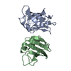 4uznC  4v2xC  4uzp C: citing same article ( |
|---|---|
| Similar structure data |


-Links
 PDBj
PDBj- Assembly
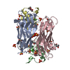
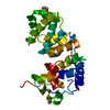
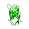
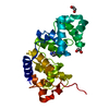
| Deposited unit | 
| ||||||||||||
|---|---|---|---|---|---|---|---|---|---|---|---|---|---|
| 1 |
| ||||||||||||
| Unit cell |
| ||||||||||||
| Components on special symmetry positions |
|
-Components
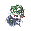
| #1: Protein | Mass: 14514.273 Da / Num. of mol.: 2 Fragment: CARBOHYDRATE BINDING MODULE FAMILY 46, UNP RESIDUES 457-563 Source method: isolated from a genetically manipulated source Details: SELENOMETHIONYLATED DERIVATIVE / Source: (gene. exp.) BACILLUS HALODURANS (bacteria) / Production host: ESCHERICHIA COLI BL21 (bacteria) / References: UniProt: Q9KF82#2: Chemical | ChemComp-SO4 / | Sulfate  Mass: 96.063 Da / Num. of mol.: 1 / Source method: obtained synthetically / Formula: SO4 Mass: 96.063 Da / Num. of mol.: 1 / Source method: obtained synthetically / Formula: SO4#3: Water | ChemComp-HOH / | Water Mass: 18.015 Da / Num. of mol.: 59 / Source method: isolated from a natural source / Formula: H2O Mass: 18.015 Da / Num. of mol.: 59 / Source method: isolated from a natural source / Formula: H2ONonpolymer details | SULFATE ION (SO4): FROM CRYSTALLIS | |
|---|
-Experimental details
-Experiment
| Experiment | Method: X-RAY DIFFRACTION / Number of used crystals: 1 |
|---|
- Sample preparation
| Crystal | Density Matthews: 2.5 Å3/Da / Density % sol: 50 % / Description: NONE |
|---|---|
| Crystal grow | pH: 7 Details: 0.2 M AMMONIUM SULFATE, 30% W/V POLYETHYLENE GLYCOL 4,000, pH 7.0 |
-Data collection
| Diffraction | Mean temperature: 100 K |
|---|---|
| Diffraction source | Source: SYNCHROTRON / Site: Diamond  / Beamline: I02 / Wavelength: 0.97976 / Beamline: I02 / Wavelength: 0.97976 |
| Detector | Type: DECTRIS PILATUS 6M / Detector: PIXEL / Date: May 6, 2013 |
| Radiation | Protocol: SINGLE WAVELENGTH / Monochromatic (M) / Laue (L): M / Scattering type: x-ray |
| Radiation wavelength | Wavelength: 0.97976 Å / Relative weight: 1 |
| Reflection | Resolution: 2.3→85.45 Å / Num. obs: 12870 / % possible obs: 100 % / Observed criterion σ(I): 0 / Redundancy: 19.1 % / Rmerge(I) obs: 0.12 / Net I/σ(I): 17.1 |
| Reflection shell | Resolution: 2.3→2.38 Å / Redundancy: 7.5 % / Rmerge(I) obs: 0.8 / Mean I/σ(I) obs: 1.3 / % possible all: 99.6 |
- Processing
| Software |
| ||||||||||||||||||||||||||||||||||||||||||||||||||||||||||||||||||||||||||||||||||||||||||||||||||||||||||||||||||||||||||||||||||||||||||||||||||||||||||||||||||||||||||||||||||||||
|---|---|---|---|---|---|---|---|---|---|---|---|---|---|---|---|---|---|---|---|---|---|---|---|---|---|---|---|---|---|---|---|---|---|---|---|---|---|---|---|---|---|---|---|---|---|---|---|---|---|---|---|---|---|---|---|---|---|---|---|---|---|---|---|---|---|---|---|---|---|---|---|---|---|---|---|---|---|---|---|---|---|---|---|---|---|---|---|---|---|---|---|---|---|---|---|---|---|---|---|---|---|---|---|---|---|---|---|---|---|---|---|---|---|---|---|---|---|---|---|---|---|---|---|---|---|---|---|---|---|---|---|---|---|---|---|---|---|---|---|---|---|---|---|---|---|---|---|---|---|---|---|---|---|---|---|---|---|---|---|---|---|---|---|---|---|---|---|---|---|---|---|---|---|---|---|---|---|---|---|---|---|---|---|
| Refinement | Method to determine structure: SAD Starting model: NONE Resolution: 2.3→85.45 Å / Cor.coef. Fo:Fc: 0.945 / Cor.coef. Fo:Fc free: 0.92 / SU B: 17.115 / SU ML: 0.202 / Cross valid method: THROUGHOUT / ESU R: 0.276 / ESU R Free: 0.225 / Stereochemistry target values: MAXIMUM LIKELIHOOD Details: HYDROGENS HAVE BEEN ADDED IN THE RIDING POSITIONS. U VALUES WITH TLS ADDED. THE COORDINATE FILE WA REFINED BY PDB_REDO IN THE PENULTIMATE ROOUND OF REFINEMENT
| ||||||||||||||||||||||||||||||||||||||||||||||||||||||||||||||||||||||||||||||||||||||||||||||||||||||||||||||||||||||||||||||||||||||||||||||||||||||||||||||||||||||||||||||||||||||
| Solvent computation | Ion probe radii: 0.8 Å / Shrinkage radii: 0.8 Å / VDW probe radii: 1 Å / Solvent model: MASK | ||||||||||||||||||||||||||||||||||||||||||||||||||||||||||||||||||||||||||||||||||||||||||||||||||||||||||||||||||||||||||||||||||||||||||||||||||||||||||||||||||||||||||||||||||||||
| Displacement parameters | Biso mean: 55.834 Å2
| ||||||||||||||||||||||||||||||||||||||||||||||||||||||||||||||||||||||||||||||||||||||||||||||||||||||||||||||||||||||||||||||||||||||||||||||||||||||||||||||||||||||||||||||||||||||
| Refinement step | Cycle: LAST / Resolution: 2.3→85.45 Å
| ||||||||||||||||||||||||||||||||||||||||||||||||||||||||||||||||||||||||||||||||||||||||||||||||||||||||||||||||||||||||||||||||||||||||||||||||||||||||||||||||||||||||||||||||||||||
| Refine LS restraints |
|