 Movie
Movie Controller


+ Open data

- Basic information
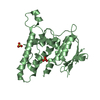
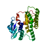
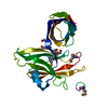
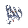
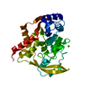
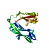
| Entry | Database: PDB / ID: 4usl | ||||||
|---|---|---|---|---|---|---|---|
| Title | The X-ray structure of calcium bound human sorcin | ||||||
 Components Components | (SORCIN SRI (gene)) x 2 SRI (gene)) x 2 | ||||||
 Keywords Keywords | METAL BINDING PROTEIN / PENTA EF-HANDS CALCIUM BINDING PROTEIN / ENDOPLASMIC RETICULUM STRESS | ||||||
| Function / homology |  Function and homology information Function and homology informationregulation of relaxation of muscle / regulation of high voltage-gated calcium channel activity / regulation of cell communication by electrical coupling / regulation of striated muscle contraction / negative regulation of cardiac muscle contraction / regulation of cardiac muscle cell contraction / Sodium/Calcium exchangers / regulation of heart contraction / muscle organ development / Reduction of cytosolic Ca++ levels ...regulation of relaxation of muscle / regulation of high voltage-gated calcium channel activity / regulation of cell communication by electrical coupling / regulation of striated muscle contraction / negative regulation of cardiac muscle contraction / regulation of cardiac muscle cell contraction / Sodium/Calcium exchangers / regulation of heart contraction / muscle organ development / Reduction of cytosolic Ca++ levels / action potential / negative regulation of heart rate / regulation of cell communication by electrical coupling involved in cardiac conduction / positive regulation of insulin secretion involved in cellular response to glucose stimulus / regulation of calcium ion transport / Ion transport by P-type ATPases / intracellular sequestering of iron ion / calcium channel regulator activity / negative regulation of ryanodine-sensitive calcium-release channel activity / regulation of release of sequestered calcium ion into cytosol by sarcoplasmic reticulum / Ion homeostasis / T-tubule / sarcoplasmic reticulum membrane / positive regulation of release of sequestered calcium ion into cytosol / sarcoplasmic reticulum / Stimuli-sensing channels / Z disc / calcium ion transport / heart development / DNA-binding transcription factor binding / protease binding / transmembrane transporter binding / protein heterodimerization activity / signaling receptor binding / calcium ion binding / endoplasmic reticulum membrane / signal transduction / extracellular exosome / nucleoplasm / membrane / identical protein binding / cytosol / cytoplasmSimilarity search - Function | ||||||
| Biological species |  HOMO SAPIENS (human) HOMO SAPIENS (human) | ||||||
| Method | X-RAY DIFFRACTION / SYNCHROTRON / MOLECULAR REPLACEMENT / Resolution: 1.65 Å | ||||||
 Authors Authors | Ilari, A. / Fiorillo, A. / Colotti, G. | ||||||
 Citation Citation | Journal: Sci.Rep. / Year: 2015 Title: Structural Basis of Sorcin-Mediated Calcium-Dependent Signal Transduction. Authors: Ilari, A. / Fiorillo, A. / Poser, E. / Lalioti, V.S. / Sundell, G.N. / Ivarsson, Y. / Genovese, I. / Colotti, G. | ||||||
| History |
|
- Structure visualization
| Structure viewer | Molecule: MolmilJmol/JSmol |
|---|
- Downloads & links
-Download
| PDBx/mmCIF format | 4usl.cif.gz | 54.2 KB | Display | PDBx/mmCIF format |
|---|---|---|---|---|
| PDB format | pdb4usl.ent.gz | 38.4 KB | Display | PDB format |
| PDBx/mmJSON format | 4usl.json.gz | Tree view | PDBx/mmJSON format | |
| Others |  Other downloads Other downloads |
-Validation report
| Arichive directory | https://data.pdbj.org/pub/pdb/validation_reports/us/4uslftp://data.pdbj.org/pub/pdb/validation_reports/us/4usl | HTTPS FTP |
|---|
-Related structure data
| Related structure data | 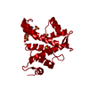 4upgC  1juoS S: Starting model for refinement C: citing same article ( |
|---|---|
| Similar structure data |

-Links
 PDBj
PDBj


- Assembly
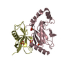
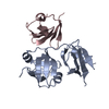
| Deposited unit | 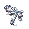
| |||||||||
|---|---|---|---|---|---|---|---|---|---|---|
| 1 |
| |||||||||
| Unit cell |
| |||||||||
| Components on special symmetry positions |
|
-Components
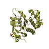
-Protein / Protein/peptide , 2 types, 2 molecules AD
| #1: Protein | SRI (gene) / 22 KDA PROTEIN / CP-22 / CP22 / V19 / SOLUBLE RESISTANCE RELATED Mass: 21695.336 Da / Num. of mol.: 1 Source method: isolated from a genetically manipulated source Source: (gene. exp.) HOMO SAPIENS (human) / Production host:  ESCHERICHIA COLI (E. coli) / Strain (production host): BL21(DE3) / References: UniProt: P30626 ESCHERICHIA COLI (E. coli) / Strain (production host): BL21(DE3) / References: UniProt: P30626 |
|---|---|
| #2: Protein/peptide | SRI (gene) / 22 KDA PROTEIN / CP-22 / CP22 / V19 Mass: 2996.209 Da / Num. of mol.: 1 / Fragment: RESIDUES 1-32 Source method: isolated from a genetically manipulated source Source: (gene. exp.) HOMO SAPIENS (human) / Production host: ESCHERICHIA COLI (E. coli) / Strain (production host): BL21(DE3) / References: UniProt: P30626 |
-Non-polymers , 4 types, 130 molecules 


| #3: Chemical |  Mass: 40.078 Da / Num. of mol.: 3 / Source method: obtained synthetically / Formula: Ca Mass: 40.078 Da / Num. of mol.: 3 / Source method: obtained synthetically / Formula: Ca#4: Chemical | ChemComp-SO4 / | Sulfate Mass: 96.063 Da / Num. of mol.: 1 / Source method: obtained synthetically / Formula: SO4 Mass: 96.063 Da / Num. of mol.: 1 / Source method: obtained synthetically / Formula: SO4#5: Chemical | Diethylene glycol Mass: 106.120 Da / Num. of mol.: 2 / Source method: obtained synthetically / Formula: C4H10O3 Mass: 106.120 Da / Num. of mol.: 2 / Source method: obtained synthetically / Formula: C4H10O3#6: Water | ChemComp-HOH / | WaterMass: 18.015 Da / Num. of mol.: 124 / Source method: isolated from a natural source / Formula: H2O |
|---|
-Experimental details
-Experiment
| Experiment | Method: X-RAY DIFFRACTION / Number of used crystals: 1 |
|---|
- Sample preparation
| Crystal | Density Matthews: 1.97 Å3/Da / Density % sol: 37.18 % / Description: NONE |
|---|---|
| Crystal grow | pH: 8.5 Details: PEG 3350 25% W/V, LISO4 0.5 M, 0.1 M TRIS, PH=8.5, CACL2 0.005M |
-Data collection
| Diffraction | Mean temperature: 100 K |
|---|---|
| Diffraction source | Source: SYNCHROTRON / Site: ELETTRA  / Beamline: 5.2R / Wavelength: 1 / Beamline: 5.2R / Wavelength: 1 |
| Detector | Type: DECTRIS PILATUS 2M / Detector: PIXEL |
| Radiation | Protocol: SINGLE WAVELENGTH / Monochromatic (M) / Laue (L): M / Scattering type: x-ray |
| Radiation wavelength | Wavelength: 1 Å / Relative weight: 1 |
| Reflection | Resolution: 1.65→50 Å / Num. obs: 20590 / % possible obs: 99.5 % / Observed criterion σ(I): 1 / Redundancy: 3.4 % / Rmerge(I) obs: 0.033 / Net I/σ(I): 21.57 |
| Reflection shell | Resolution: 1.65→1.69 Å / Redundancy: 3.3 % / Rmerge(I) obs: 0.55 / Mean I/σ(I) obs: 3 / % possible all: 99.6 |
- Processing
| Software |
| ||||||||||||||||||||||||||||||||||||||||||||||||||||||||||||||||||||||||||||||||||||||||||||||||||||||||||||||||||||||||||||||||||||||||||||||||||||||||||||||||||||||||||||||||||||||
|---|---|---|---|---|---|---|---|---|---|---|---|---|---|---|---|---|---|---|---|---|---|---|---|---|---|---|---|---|---|---|---|---|---|---|---|---|---|---|---|---|---|---|---|---|---|---|---|---|---|---|---|---|---|---|---|---|---|---|---|---|---|---|---|---|---|---|---|---|---|---|---|---|---|---|---|---|---|---|---|---|---|---|---|---|---|---|---|---|---|---|---|---|---|---|---|---|---|---|---|---|---|---|---|---|---|---|---|---|---|---|---|---|---|---|---|---|---|---|---|---|---|---|---|---|---|---|---|---|---|---|---|---|---|---|---|---|---|---|---|---|---|---|---|---|---|---|---|---|---|---|---|---|---|---|---|---|---|---|---|---|---|---|---|---|---|---|---|---|---|---|---|---|---|---|---|---|---|---|---|---|---|---|---|
| Refinement | Method to determine structure: MOLECULAR REPLACEMENT Starting model: PDB ENTRY 1JUO Resolution: 1.65→37.34 Å / Cor.coef. Fo:Fc: 0.961 / Cor.coef. Fo:Fc free: 0.95 / SU B: 2.248 / SU ML: 0.076 / Cross valid method: THROUGHOUT / ESU R: 0.108 / ESU R Free: 0.103 / Stereochemistry target values: MAXIMUM LIKELIHOOD / Details: HYDROGENS HAVE BEEN ADDED IN THE RIDING POSITIONS.
| ||||||||||||||||||||||||||||||||||||||||||||||||||||||||||||||||||||||||||||||||||||||||||||||||||||||||||||||||||||||||||||||||||||||||||||||||||||||||||||||||||||||||||||||||||||||
| Solvent computation | Ion probe radii: 0.8 Å / Shrinkage radii: 0.8 Å / VDW probe radii: 1.2 Å / Solvent model: MASK | ||||||||||||||||||||||||||||||||||||||||||||||||||||||||||||||||||||||||||||||||||||||||||||||||||||||||||||||||||||||||||||||||||||||||||||||||||||||||||||||||||||||||||||||||||||||
| Displacement parameters | Biso mean: 26.993 Å2
| ||||||||||||||||||||||||||||||||||||||||||||||||||||||||||||||||||||||||||||||||||||||||||||||||||||||||||||||||||||||||||||||||||||||||||||||||||||||||||||||||||||||||||||||||||||||
| Refinement step | Cycle: LAST / Resolution: 1.65→37.34 Å
| ||||||||||||||||||||||||||||||||||||||||||||||||||||||||||||||||||||||||||||||||||||||||||||||||||||||||||||||||||||||||||||||||||||||||||||||||||||||||||||||||||||||||||||||||||||||
| Refine LS restraints |
|