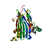
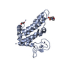
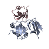
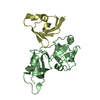
[English] 日本語
Yorodumi
- PDB-4kdi: Crystal structure of p97/VCP N in complex with OTU1 UBXL -

+
Open data


ID or keywords:

Loading...

-
Basic information

Entry
Database: PDB / ID: 4kdi
TitleCrystal structure of p97/VCP N in complex with OTU1 UBXL
Components
  • Transitional endoplasmic reticulum ATPase
  • Ubiquitin thioesterase OTU1
KeywordsSIGNALING PROTEIN/HYDROLASE / AAA ATPase / PROTEIN-PROTEIN COMPLEX / UBX-like domain / PROTEIN BINDING / beta-barrel / beta-grasp / SIGNALING PROTEIN-HYDROLASE complex
Function / homology
Function and homology information


Ovarian tumor domain proteases / positive regulation of Lys63-specific deubiquitinase activity / flavin adenine dinucleotide catabolic process / positive regulation of oxidative phosphorylation / VCP-NSFL1C complex / endosome to lysosome transport via multivesicular body sorting pathway / protein-DNA covalent cross-linking repair / endoplasmic reticulum stress-induced pre-emptive quality control / cellular response to arsenite ion / Derlin-1 retrotranslocation complex ...Ovarian tumor domain proteases / positive regulation of Lys63-specific deubiquitinase activity / flavin adenine dinucleotide catabolic process / positive regulation of oxidative phosphorylation / VCP-NSFL1C complex / endosome to lysosome transport via multivesicular body sorting pathway / protein-DNA covalent cross-linking repair / endoplasmic reticulum stress-induced pre-emptive quality control / cellular response to arsenite ion / Derlin-1 retrotranslocation complex / BAT3 complex binding / positive regulation of protein K63-linked deubiquitination / deubiquitinase activator activity / mitotic spindle disassembly / VCP-NPL4-UFD1 AAA ATPase complex / aggresome assembly / regulation of protein localization to chromatin / vesicle-fusing ATPase / NADH metabolic process / cellular response to misfolded protein / stress granule disassembly / K48-linked polyubiquitin modification-dependent protein binding / positive regulation of mitochondrial membrane potential / negative regulation of protein localization to chromatin / ubiquitin-modified protein reader activity / retrograde protein transport, ER to cytosol / regulation of aerobic respiration / ATPase complex / regulation of synapse organization / ubiquitin-specific protease binding / positive regulation of ATP biosynthetic process / : / protein deubiquitination / ERAD pathway / ubiquitin-like protein ligase binding / MHC class I protein binding / autophagosome maturation / RHOH GTPase cycle / polyubiquitin modification-dependent protein binding / HSF1 activation / translesion synthesis / Protein methylation / endoplasmic reticulum to Golgi vesicle-mediated transport / interstrand cross-link repair / negative regulation of smoothened signaling pathway / ATP metabolic process / Attachment and Entry / endoplasmic reticulum unfolded protein response / viral genome replication / lipid droplet / proteasome complex / proteasomal protein catabolic process / Josephin domain DUBs / N-glycan trimming in the ER and Calnexin/Calreticulin cycle / Hh mutants are degraded by ERAD / Defective CFTR causes cystic fibrosis / Hedgehog ligand biogenesis / macroautophagy / Translesion Synthesis by POLH / positive regulation of protein-containing complex assembly / ABC-family proteins mediated transport / establishment of protein localization / ADP binding / autophagy / Aggrephagy / cytoplasmic stress granule / positive regulation of non-canonical NF-kappaB signal transduction / positive regulation of protein catabolic process / activation of cysteine-type endopeptidase activity involved in apoptotic process / KEAP1-NFE2L2 pathway / azurophil granule lumen / double-strand break repair / positive regulation of canonical Wnt signaling pathway / Ovarian tumor domain proteases / positive regulation of proteasomal ubiquitin-dependent protein catabolic process / E3 ubiquitin ligases ubiquitinate target proteins / site of double-strand break / Neddylation / cellular response to heat / ubiquitin-dependent protein catabolic process / regulation of apoptotic process / protein phosphatase binding / proteasome-mediated ubiquitin-dependent protein catabolic process / secretory granule lumen / ubiquitinyl hydrolase 1 / ficolin-1-rich granule lumen / cysteine-type deubiquitinase activity / Attachment and Entry / protein ubiquitination / protein domain specific binding / intracellular membrane-bounded organelle / DNA repair / glutamatergic synapse / lipid binding / DNA damage response / ubiquitin protein ligase binding / Neutrophil degranulation / endoplasmic reticulum membrane / regulation of DNA-templated transcription / perinuclear region of cytoplasm
Similarity search - Function
: / OTU1, UBXL domain / Vcp-like ATPase; Chain A, domain 2 - #10 / OTU-like cysteine protease / Vcp-like ATPase; Chain A, domain 2 / OTU domain / OTU domain profile. / Barwin-like endoglucanases - #20 / AAA ATPase, CDC48 family / Barwin-like endoglucanases ...: / OTU1, UBXL domain / Vcp-like ATPase; Chain A, domain 2 - #10 / OTU-like cysteine protease / Vcp-like ATPase; Chain A, domain 2 / OTU domain / OTU domain profile. / Barwin-like endoglucanases - #20 / AAA ATPase, CDC48 family / Barwin-like endoglucanases / Cell division protein 48 (CDC48), N-terminal domain / CDC48, N-terminal subdomain / Cell division protein 48 (CDC48) N-terminal domain / CDC48, domain 2 / Cell division protein 48 (CDC48), domain 2 / Cell division protein 48 (CDC48) domain 2 / CDC48 domain 2-like superfamily / Aspartate decarboxylase-like domain superfamily / AAA ATPase, AAA+ lid domain / AAA+ lid domain / ATPase, AAA-type, conserved site / AAA-protein family signature. / Phosphatidylinositol 3-kinase Catalytic Subunit; Chain A, domain 1 / Zinc finger C2H2 type domain signature. / Zinc finger C2H2-type / Papain-like cysteine peptidase superfamily / ATPase family associated with various cellular activities (AAA) / ATPase, AAA-type, core / Ubiquitin-like (UB roll) / ATPases associated with a variety of cellular activities / AAA+ ATPase domain / Roll / Beta Barrel / P-loop containing nucleoside triphosphate hydrolase / Mainly Beta / Alpha Beta
Similarity search - Domain/homology
Ubiquitin thioesterase OTU1 / Transitional endoplasmic reticulum ATPase
Similarity search - Component
Biological speciesHomo sapiens (human)
Saccharomyces cerevisiae (brewer's yeast)
MethodX-RAY DIFFRACTION / SYNCHROTRON / MOLECULAR REPLACEMENT / Resolution: 1.86 Å
AuthorsKim, S.J. / Kim, E.E.
CitationJournal: J.Biol.Chem. / Year: 2014
Title: Structural Basis for Ovarian Tumor Domain-containing Protein 1 (OTU1) Binding to p97/Valosin-containing Protein (VCP).
Authors: Kim, S.J. / Cho, J. / Song, E.J. / Kim, S.J. / Kim, H.M. / Lee, K.E. / Suh, S.W. / Kim, E.E.
History
DepositionApr 25, 2013Deposition site: RCSB / Processing site: RCSB
Revision 1.0Mar 19, 2014Provider: repository / Type: Initial release
Revision 1.1May 21, 2014Group: Database references
Revision 1.2Feb 28, 2024Group: Data collection / Database references
Category: chem_comp_atom / chem_comp_bond ...chem_comp_atom / chem_comp_bond / database_2 / struct_ref_seq_dif
Item: _database_2.pdbx_DOI / _database_2.pdbx_database_accession / _struct_ref_seq_dif.details

-
Structure visualization

Structure viewerMolecule:
MolmilJmol/JSmol

Downloads & links

-
Assembly

Deposited unit
A: Transitional endoplasmic reticulum ATPase
B: Transitional endoplasmic reticulum ATPase
C: Ubiquitin thioesterase OTU1
D: Ubiquitin thioesterase OTU1


Theoretical massNumber of molelcules
Total (without water)63,0004
Polymers63,0004
Non-polymers00
Water3,567198
1
A: Transitional endoplasmic reticulum ATPase
C: Ubiquitin thioesterase OTU1


Theoretical massNumber of molelcules
Total (without water)31,5002
Polymers31,5002
Non-polymers00
Water362
TypeNameSymmetry operationNumber
identity operation1_555x,y,z1
2
B: Transitional endoplasmic reticulum ATPase
D: Ubiquitin thioesterase OTU1


Theoretical massNumber of molelcules
Total (without water)31,5002
Polymers31,5002
Non-polymers00
Water362
TypeNameSymmetry operationNumber
identity operation1_555x,y,z1
Unit cell
Length a, b, c (Å)42.819, 88.609, 143.535
Angle α, β, γ (deg.)90.00, 90.00, 90.00
Int Tables number19
Space group name H-MP212121

-
Components

#1: Protein Transitional endoplasmic reticulum ATPase / TER ATPase / 15S Mg(2+)-ATPase p97 subunit / Valosin-containing protein / VCP


Mass: 21806.027 Da / Num. of mol.: 2 / Fragment: N domain (Residues 21-185)
Source method: isolated from a genetically manipulated source
Source: (gene. exp.) Homo sapiens (human) / Gene: VCP / Plasmid: pET28a / Production host: Escherichia coli (E. coli) / Strain (production host): Rosetta (DE3) / References: UniProt: P55072, vesicle-fusing ATPase
#2: Protein Ubiquitin thioesterase OTU1 / OTU domain-containing protein 1


Mass: 9693.965 Da / Num. of mol.: 2 / Fragment: UBX-like domain (Residues 1-73)
Source method: isolated from a genetically manipulated source
Source: (gene. exp.) Saccharomyces cerevisiae (brewer's yeast)
Strain: ATCC 204508 / S288c / Gene: OTU1, YFL044C / Plasmid: pET28a / Production host: Escherichia coli (E. coli) / Strain (production host): Rosetta (DE3) / References: UniProt: P43558, ubiquitinyl hydrolase 1
#3: Water ChemComp-HOH / water / Water


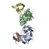
Mass: 18.015 Da / Num. of mol.: 198 / Source method: isolated from a natural source / Formula: H2O

-
Experimental details

-
Experiment

ExperimentMethod: X-RAY DIFFRACTION / Number of used crystals: 1

-
Sample preparation

CrystalDensity Matthews: 2.16 Å3/Da / Density % sol: 43.08 %
Crystal growTemperature: 295 K / Method: vapor diffusion, hanging drop / pH: 5
Details: 20% PEG 2000, 10% Tacsimate, pH 5.0, VAPOR DIFFUSION, HANGING DROP, temperature 295.0K

-
Data collection

DiffractionMean temperature: 100 K
Diffraction sourceSource: SYNCHROTRON / Site: PAL/PLS / Beamline: 4A / Wavelength: 0.97951 Å
DetectorType: ADSC QUANTUM 315r / Detector: CCD / Date: Jul 20, 2010
RadiationMonochromator: Si(111) / Protocol: SINGLE WAVELENGTH / Monochromatic (M) / Laue (L): M / Scattering type: x-ray
Radiation wavelengthWavelength: 0.97951 Å / Relative weight: 1
ReflectionResolution: 1.86→50 Å / Num. obs: 46883 / % possible obs: 96.5 % / Observed criterion σ(F): 0 / Observed criterion σ(I): -3 / Redundancy: 8.5 % / Rmerge(I) obs: 0.089 / Rsym value: 0.089 / Net I/σ(I): 25
Reflection shellResolution: 1.86→1.93 Å / Redundancy: 4.9 % / Rmerge(I) obs: 0.385 / Mean I/σ(I) obs: 2.631 / Rsym value: 0.385 / % possible all: 88.9

-
Processing

Software
NameVersionClassification
HKL-2000data collection
PHASERphasing
REFMAC5.5.0109refinement
HKL-2000data reduction
HKL-2000data scaling
RefinementMethod to determine structure: MOLECULAR REPLACEMENT / Resolution: 1.86→42.34 Å / Cor.coef. Fo:Fc: 0.955 / Cor.coef. Fo:Fc free: 0.932 / SU B: 10.375 / SU ML: 0.132 / Cross valid method: THROUGHOUT / ESU R: 0.154 / ESU R Free: 0.148 / Stereochemistry target values: MAXIMUM LIKELIHOOD / Details: HYDROGENS HAVE BEEN ADDED IN THE RIDING POSITIONS
RfactorNum. reflection% reflectionSelection details
Rfree0.25677 2284 5.1 %RANDOM
Rwork0.21367 ---
obs0.21584 42901 96.64 %-
Solvent computationIon probe radii: 0.8 Å / Shrinkage radii: 0.8 Å / VDW probe radii: 1.4 Å / Solvent model: MASK
Displacement parametersBiso mean: 46.039 Å2
Baniso -1Baniso -2Baniso -3
1-1.12 Å20 Å20 Å2
2--1.19 Å20 Å2
3----2.31 Å2
Refinement stepCycle: LAST / Resolution: 1.86→42.34 Å
ProteinNucleic acidLigandSolventTotal
Num. atoms3665 0 0 198 3863
Refine LS restraints
Refine-IDTypeDev idealDev ideal targetNumber
X-RAY DIFFRACTIONr_bond_refined_d0.0230.0223713
X-RAY DIFFRACTIONr_angle_refined_deg1.9261.9875014
X-RAY DIFFRACTIONr_dihedral_angle_1_deg6.8265457
X-RAY DIFFRACTIONr_dihedral_angle_2_deg37.53624.368174
X-RAY DIFFRACTIONr_dihedral_angle_3_deg17.51215701
X-RAY DIFFRACTIONr_dihedral_angle_4_deg16.1821534
X-RAY DIFFRACTIONr_chiral_restr0.1350.2586
X-RAY DIFFRACTIONr_gen_planes_refined0.0090.0212750
X-RAY DIFFRACTIONr_mcbond_it1.351.52303
X-RAY DIFFRACTIONr_mcangle_it2.31323763
X-RAY DIFFRACTIONr_scbond_it3.31631410
X-RAY DIFFRACTIONr_scangle_it5.4694.51251
LS refinement shellResolution: 1.861→1.909 Å / Total num. of bins used: 20
RfactorNum. reflection% reflection
Rfree0.413 145 -
Rwork0.401 2885 -
obs--90.42 %
Refinement TLS params.

Method: refined / Refine-ID: X-RAY DIFFRACTION

IDL112)L122)L132)L222)L232)L332)S11 (Å °)S12 (Å °)S13 (Å °)S21 (Å °)S22 (Å °)S23 (Å °)S31 (Å °)S32 (Å °)S33 (Å °)T112)T122)T132)T222)T232)T332)Origin x (Å)Origin y (Å)Origin z (Å)
10.6516-0.52890.73340.7117-0.3691.1473-0.04510.00360.0116-0.0988-0.0254-0.0821-0.11410.00530.07050.1437-0.01010.03010.1310.06220.146520.157544.839737.0083
20.3215-0.15140.07150.08390.01492.90890.1688-0.0031-0.0633-0.0635-0.00040.06210.42340.3273-0.16840.23060.1375-0.0330.1872-0.03240.13261.786650.15175.0373
32.2332-2.05311.46443.1767-2.31521.689-0.5379-0.30140.14520.66040.2545-0.3675-0.4699-0.17340.28330.41820.0712-0.1560.0535-0.02770.100617.484466.074947.9888
40.51390.112-0.08231.77420.750.3891-0.04970.07280.05460.0648-0.01510.19040.02570.05360.06480.1050.035-0.03050.2105-0.00990.1074-0.986465.6016-14.9418
Refinement TLS group
IDRefine-IDRefine TLS-IDAuth asym-IDAuth seq-ID
1X-RAY DIFFRACTION1A21 - 185
2X-RAY DIFFRACTION2B22 - 187
3X-RAY DIFFRACTION3C2 - 73
4X-RAY DIFFRACTION4D-1 - 73

+
About Yorodumi

-
News

-
Feb 9, 2022. New format data for meta-information of EMDB entries

New format data for meta-information of EMDB entries

  • Version 3 of the EMDB header file is now the official format.
  • The previous official version 1.9 will be removed from the archive.

Related info.:EMDB header

External links:wwPDB to switch to version 3 of the EMDB data model

-
Aug 12, 2020. Covid-19 info

Covid-19 info

URL: https://pdbj.org/emnavi/covid19.php

New page: Covid-19 featured information page in EM Navigator.

Related info.:Covid-19 info / Mar 5, 2020. Novel coronavirus structure data

+
Mar 5, 2020. Novel coronavirus structure data

Novel coronavirus structure data

Related info.:Yorodumi Speices / Aug 12, 2020. Covid-19 info

External links:COVID-19 featured content - PDBj / Molecule of the Month (242):Coronavirus Proteases

+
Jan 31, 2019. EMDB accession codes are about to change! (news from PDBe EMDB page)

EMDB accession codes are about to change! (news from PDBe EMDB page)

  • The allocation of 4 digits for EMDB accession codes will soon come to an end. Whilst these codes will remain in use, new EMDB accession codes will include an additional digit and will expand incrementally as the available range of codes is exhausted. The current 4-digit format prefixed with “EMD-” (i.e. EMD-XXXX) will advance to a 5-digit format (i.e. EMD-XXXXX), and so on. It is currently estimated that the 4-digit codes will be depleted around Spring 2019, at which point the 5-digit format will come into force.
  • The EM Navigator/Yorodumi systems omit the EMD- prefix.

Related info.:Q: What is EMD? / ID/Accession-code notation in Yorodumi/EM Navigator

External links:EMDB Accession Codes are Changing Soon! / Contact to PDBj

+
Jul 12, 2017. Major update of PDB

Major update of PDB

  • wwPDB released updated PDB data conforming to the new PDBx/mmCIF dictionary.
  • This is a major update changing the version number from 4 to 5, and with Remediation, in which all the entries are updated.
  • In this update, many items about electron microscopy experimental information are reorganized (e.g. em_software).
  • Now, EM Navigator and Yorodumi are based on the updated data.

External links:wwPDB Remediation / Enriched Model Files Conforming to OneDep Data Standards Now Available in the PDB FTP Archive

-
Yorodumi

Thousand views of thousand structures

  • Yorodumi is a browser for structure data from EMDB, PDB, SASBDB, etc.
  • This page is also the successor to EM Navigator detail page, and also detail information page/front-end page for Omokage search.
  • The word "yorodu" (or yorozu) is an old Japanese word meaning "ten thousand". "mi" (miru) is to see.

Related info.:EMDB / PDB / SASBDB / Comparison of 3 databanks / Yorodumi Search / Aug 31, 2016. New EM Navigator & Yorodumi / Yorodumi Papers / Jmol/JSmol / Function and homology information / Changes in new EM Navigator and Yorodumi

Read more