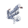
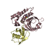
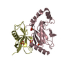
Entry Database : PDB / ID : 4x57Title Structure of an Arabidopsis E2 / Membrane-anchored Ubiquitin-fold Protein Complex Membrane-anchored ubiquitin-fold protein 3 Ubiquitin-conjugating enzyme E2 8 Keywords / / / / / / / Function / homology Biological species Arabidopsis thaliana (thale cress)Method / / / Resolution : 2.8 Å Authors Korolev, S. / Koroleva, O. / Lu, X. / Downes, B. Funding support Organization Grant number Country National Institutes of Health/National Institute of General Medical Sciences (NIH/NIGMS) GM096279
Journal : To Be Published Title : Structure of an Arabidopsis E2 / Membrane-anchored Ubiquitin-fold ProteinComplexAuthors : Korolev, S. / Koroleva, O. / Lu, X. / Downes, B. History Deposition Dec 4, 2014 Deposition site / Processing site Revision 1.0 Jan 20, 2016 Provider / Type Revision 1.1 Sep 13, 2017 Group / Derived calculations / Category / pdbx_struct_oper_listItem / _pdbx_struct_oper_list.symmetry_operationRevision 1.2 Dec 25, 2019 Group / Category / Item Revision 1.3 Sep 27, 2023 Group / Database references / Refinement descriptionCategory chem_comp_atom / chem_comp_bond ... chem_comp_atom / chem_comp_bond / database_2 / pdbx_initial_refinement_model / struct_ncs_dom_lim Item _database_2.pdbx_DOI / _database_2.pdbx_database_accession ... _database_2.pdbx_DOI / _database_2.pdbx_database_accession / _struct_ncs_dom_lim.beg_auth_comp_id / _struct_ncs_dom_lim.beg_label_asym_id / _struct_ncs_dom_lim.beg_label_comp_id / _struct_ncs_dom_lim.beg_label_seq_id / _struct_ncs_dom_lim.end_auth_comp_id / _struct_ncs_dom_lim.end_label_asym_id / _struct_ncs_dom_lim.end_label_comp_id / _struct_ncs_dom_lim.end_label_seq_id
Show all Show less
 Movie
Movie Controller

 Yorodumi
 Open data
 Basic information
 Components
 Keywords
 Ubiquitin / Ubconjugating (E2) enzymes / Membrane anchored / Ubiquitin-fold protein 3 / MUB3 / E1:E2 Complex / ligase-protein binding complex
 Function and homology information
 Authors
 United States, 1items
  Citation
 Structure visualization
 Downloads & links
 Other downloads
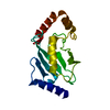


 PDBj
PDBj

 Assembly


 Mass: 96.063 Da / Num. of mol.: 6 / Source method: obtained synthetically / Formula: SO4
 Sample preparation
 Processing