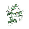
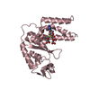
B: DNA (5'-D(*DTP*DTP*DTP*DAP*DAP*DCP*DAP*DGP*DTP*DTP*DAP*DAP*DGP*(BRU)P*DAP*DT)-3') C: DNA (5'-D(*DAP*DAP*(BRU)P*DAP*DCP*DTP*DTP*DAP*DAP*DCP*DTP*DGP*DTP*DTP*DAP*DA)-3') A: Accessory gene regulator protein A hetero molecules
Resolution: 1.6→1.66 Å / Redundancy: 3.8 % / Rmerge(I) obs: 0.155 / Mean I/σ(I) obs: 5.7 / % possible all: 97.3
-
Processing
Software
Name
Version
Classification
REFMAC
5.2.0019
refinement
ADSC
Quantum
datacollection
HKL-2000
datareduction
HKL-2000
datascaling
SHARP
phasing
Refinement
Method to determine structure: SAD / Resolution: 1.6→17.3 Å / Cor.coef. Fo:Fc: 0.952 / Cor.coef. Fo:Fc free: 0.94 / SU B: 1.717 / SU ML: 0.056 / Cross valid method: THROUGHOUT / σ(F): 0 / ESU R: 0.093 / ESU R Free: 0.091 Stereochemistry target values: MAXIMUM LIKELIHOOD WITH PHASES Details: HYDROGENS HAVE BEEN ADDED IN THE RIDING POSITIONS
Rfactor
Num. reflection
% reflection
Selection details
Rfree
0.22066
1485
5.1 %
RANDOM
Rwork
0.19534
-
-
-
all
0.1961
29714
-
-
obs
0.19659
27747
98.35 %
-
Solvent computation
Ion probe radii: 0.8 Å / Shrinkage radii: 0.8 Å / VDW probe radii: 1.2 Å / Solvent model: MASK
Displacement parameters
Biso mean: 19.945 Å2
Baniso -1
Baniso -2
Baniso -3
1-
0 Å2
0 Å2
0 Å2
2-
-
0 Å2
0 Å2
3-
-
-
0 Å2
Refinement step
Cycle: LAST / Resolution: 1.6→17.3 Å
Protein
Nucleic acid
Ligand
Solvent
Total
Num. atoms
872
650
2
223
1747
Refine LS restraints
Refine-ID
Type
Dev ideal
Dev ideal target
Number
X-RAY DIFFRACTION
r_bond_refined_d
0.007
0.021
1617
X-RAY DIFFRACTION
r_angle_refined_deg
1.381
2.445
2313
X-RAY DIFFRACTION
r_dihedral_angle_1_deg
5.998
5
104
X-RAY DIFFRACTION
r_dihedral_angle_2_deg
37.564
23.654
52
X-RAY DIFFRACTION
r_dihedral_angle_3_deg
11.461
15
169
X-RAY DIFFRACTION
r_dihedral_angle_4_deg
13.397
15
8
X-RAY DIFFRACTION
r_chiral_restr
0.073
0.2
250
X-RAY DIFFRACTION
r_gen_planes_refined
0.005
0.02
1019
X-RAY DIFFRACTION
r_nbd_refined
0.187
0.2
583
X-RAY DIFFRACTION
r_nbtor_refined
0.302
0.2
1021
X-RAY DIFFRACTION
r_xyhbond_nbd_refined
0.374
0.2
189
X-RAY DIFFRACTION
r_metal_ion_refined
0.023
0.2
2
X-RAY DIFFRACTION
r_symmetry_vdw_refined
0.222
0.2
54
X-RAY DIFFRACTION
r_symmetry_hbond_refined
0.155
0.2
34
X-RAY DIFFRACTION
r_mcbond_it
0.768
1.5
532
X-RAY DIFFRACTION
r_mcangle_it
1.216
2
839
X-RAY DIFFRACTION
r_scbond_it
1.364
3
1440
X-RAY DIFFRACTION
r_scangle_it
1.996
4.5
1473
LS refinement shell
Resolution: 1.6→1.641 Å / Total num. of bins used: 20
Rfactor
Num. reflection
% reflection
Rfree
0.256
112
-
Rwork
0.213
1983
-
obs
-
-
97.26 %
+
About Yorodumi
-
News
-
Feb 9, 2022. New format data for meta-information of EMDB entries
New format data for meta-information of EMDB entries
Version 3 of the EMDB header file is now the official format.
The previous official version 1.9 will be removed from the archive.
In the structure databanks used in Yorodumi, some data are registered as the other names, "COVID-19 virus" and "2019-nCoV". Here are the details of the virus and the list of structure data.
Jan 31, 2019. EMDB accession codes are about to change! (news from PDBe EMDB page)
EMDB accession codes are about to change! (news from PDBe EMDB page)
The allocation of 4 digits for EMDB accession codes will soon come to an end. Whilst these codes will remain in use, new EMDB accession codes will include an additional digit and will expand incrementally as the available range of codes is exhausted. The current 4-digit format prefixed with “EMD-” (i.e. EMD-XXXX) will advance to a 5-digit format (i.e. EMD-XXXXX), and so on. It is currently estimated that the 4-digit codes will be depleted around Spring 2019, at which point the 5-digit format will come into force.
The EM Navigator/Yorodumi systems omit the EMD- prefix.
Related info.:Q: What is EMD? / ID/Accession-code notation in Yorodumi/EM Navigator
Yorodumi is a browser for structure data from EMDB, PDB, SASBDB, etc.
This page is also the successor to EM Navigator detail page, and also detail information page/front-end page for Omokage search.
The word "yorodu" (or yorozu) is an old Japanese word meaning "ten thousand". "mi" (miru) is to see.
Related info.:EMDB / PDB / SASBDB / Comparison of 3 databanks / Yorodumi Search / Aug 31, 2016. New EM Navigator & Yorodumi / Yorodumi Papers / Jmol/JSmol / Function and homology information / Changes in new EM Navigator and Yorodumi
 Movie
Movie Controller

 Yorodumi
 Open data
 Basic information
 Components
 Keywords
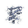
 transcription regulator / LytTR / AgrA /
  Function and homology information

 Authors
 Citation
 Structure visualization
 Downloads & links
 Other downloads
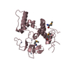


 PDBj
PDBj


 Assembly
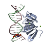

 Mass: 24.305 Da / Num. of mol.: 2 / Source method: obtained synthetically / Formula: Mg
 Mass: 18.015 Da / Num. of mol.: 223 / Source method: isolated from a natural source / Formula: H2O
 Sample preparation
 / Beamline: X4A / Wavelength: 0.9204 Å
 Processing