 Movie
Movie Controller


+ Open data

- Basic information
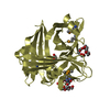
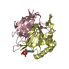
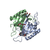
| Entry | Database: PDB / ID: 4ue0 | ||||||
|---|---|---|---|---|---|---|---|
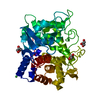
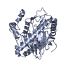
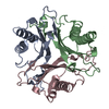
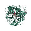
| Title | Structure of the bovine atadenovirus type 4 fibre head protein | ||||||
 Components Components | FIBER | ||||||
 Keywords Keywords | VIRAL PROTEIN | ||||||
| Function / homology |  Function and homology information Function and homology informationadhesion receptor-mediated virion attachment to host cell / viral capsid / cell adhesion / symbiont entry into host cell / host cell nucleus / nucleusSimilarity search - Function | ||||||
| Biological species |  BOVINE ADENOVIRUS 4 BOVINE ADENOVIRUS 4 | ||||||
| Method | X-RAY DIFFRACTION / SYNCHROTRON / SIRAS / Resolution: 1.17 Å | ||||||
 Authors Authors | Nguyen, T.H. / van Raaij, M.J. | ||||||
 Citation Citation | Journal: Virol. J. / Year: 2015 Title: Crystal structure of the fibre head domain of bovine adenovirus 4, a ruminant atadenovirus. Authors: Nguyen, T.H. / Vidovszky, M.Z. / Ballmann, M.Z. / Sanz-Gaitero, M. / Singh, A.K. / Harrach, B. / Benko, M. / van Raaij, M.J. | ||||||
| History |
|
- Structure visualization
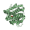
| Structure viewer | Molecule: MolmilJmol/JSmol |
|---|
- Downloads & links
-Download
| PDBx/mmCIF format | 4ue0.cif.gz | 168.4 KB | Display | PDBx/mmCIF format |
|---|---|---|---|---|
| PDB format | pdb4ue0.ent.gz | 136.5 KB | Display | PDB format |
| PDBx/mmJSON format | 4ue0.json.gz | Tree view | PDBx/mmJSON format | |
| Others |  Other downloads Other downloads |
-Validation report
| Arichive directory | https://data.pdbj.org/pub/pdb/validation_reports/ue/4ue0ftp://data.pdbj.org/pub/pdb/validation_reports/ue/4ue0 | HTTPS FTP |
|---|
-Related structure data
| Similar structure data |
|---|
-Links
 PDBj
PDBj


- Assembly
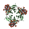
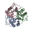
| Deposited unit | 
| ||||||||
|---|---|---|---|---|---|---|---|---|---|
| 1 |
| ||||||||
| Unit cell |
|
-Components
| #1: Protein | Mass: 13561.470 Da / Num. of mol.: 3 / Fragment: HEAD DOMAIN, RESIDUES 414-535 Source method: isolated from a genetically manipulated source Source: (gene. exp.) BOVINE ADENOVIRUS 4Description: PROTOTYPE STRAIN OF BADV-4 (CALLED THT/62) WAS ISOLATED BY ADORJAN BARTHA IN 1962. THE GENOME IN THE GENBANK IS UNDER ACCESSION NUMBER NC_002685.2. IT IS A SO CALLED REFSEQ ENTRY, ...Description: PROTOTYPE STRAIN OF BADV-4 (CALLED THT/62) WAS ISOLATED BY ADORJAN BARTHA IN 1962. THE GENOME IN THE GENBANK IS UNDER ACCESSION NUMBER NC_002685.2. IT IS A SO CALLED REFSEQ ENTRY, MEANING THAT BADV-4 IS THE REFERENCE STRAIN FOR THE BOVINE ATADENOVIRUS D SPECIES. REFERENCE BARTHA, A., ALDASY, P. (1966) FURTHER TWO SEROTYPES OF BOVINE ADENOVIRUS (SEROTYPE 4 AND 5). ACTA VET. HUNG. ACAD. SCI. 16(1)107-108. Production host:  ESCHERICHIA COLI (E. coli) / Strain (production host): BL21(DE3) / Variant (production host): JM109 / References: UniProt: Q997H2 ESCHERICHIA COLI (E. coli) / Strain (production host): BL21(DE3) / Variant (production host): JM109 / References: UniProt: Q997H2#2: Chemical | ChemComp-CO3 / | Carbonate  Mass: 60.009 Da / Num. of mol.: 1 / Source method: obtained synthetically / Formula: CO3 Mass: 60.009 Da / Num. of mol.: 1 / Source method: obtained synthetically / Formula: CO3#3: Chemical | ChemComp-SCN / | Thiocyanate  Mass: 58.082 Da / Num. of mol.: 1 / Source method: obtained synthetically / Formula: CNS Mass: 58.082 Da / Num. of mol.: 1 / Source method: obtained synthetically / Formula: CNS#4: Water | ChemComp-HOH / | Water Mass: 18.015 Da / Num. of mol.: 441 / Source method: isolated from a natural source / Formula: H2O Mass: 18.015 Da / Num. of mol.: 441 / Source method: isolated from a natural source / Formula: H2O |
|---|
-Experimental details
-Experiment
| Experiment | Method: X-RAY DIFFRACTION / Number of used crystals: 2 |
|---|
- Sample preparation
| Crystal | Density Matthews: 2.2 Å3/Da / Density % sol: 44 % / Description: NONE |
|---|---|
| Crystal grow | Details: 20% (W/V) PEG3350, 200 MM KSCN, 50 MM NACL, 10 MM TRIS-HCL PH 7.5, 5% (V/V) GLYCEROL PH range: 7.5; 7.5 |
-Data collection
| Diffraction |
| ||||||||||||||||||
|---|---|---|---|---|---|---|---|---|---|---|---|---|---|---|---|---|---|---|---|
| Diffraction source |
| ||||||||||||||||||
| Detector |
| ||||||||||||||||||
| Radiation |
| ||||||||||||||||||
| Radiation wavelength |
| ||||||||||||||||||
| Reflection | Resolution: 1.17→44.15 Å / Num. obs: 103174 / % possible obs: 91.2 % / Redundancy: 3.6 % / Biso Wilson estimate: 10.5 Å2 / Rmerge(I) obs: 0.03 / Net I/σ(I): 15.7 | ||||||||||||||||||
| Reflection shell | Resolution: 1.17→1.21 Å / Redundancy: 3.5 % / Rmerge(I) obs: 0.35 / Mean I/σ(I) obs: 2.6 / % possible all: 86.7 |


- Processing
| Software |
| ||||||||||||||||||||||||||||||||||||||||||||||||||||||||||||||||||||||||||||||||||||||||||||||||||||||||||||||||||||||||||||||||||||||||||||||||||||||||||||||||||||||||||||||||||||||
|---|---|---|---|---|---|---|---|---|---|---|---|---|---|---|---|---|---|---|---|---|---|---|---|---|---|---|---|---|---|---|---|---|---|---|---|---|---|---|---|---|---|---|---|---|---|---|---|---|---|---|---|---|---|---|---|---|---|---|---|---|---|---|---|---|---|---|---|---|---|---|---|---|---|---|---|---|---|---|---|---|---|---|---|---|---|---|---|---|---|---|---|---|---|---|---|---|---|---|---|---|---|---|---|---|---|---|---|---|---|---|---|---|---|---|---|---|---|---|---|---|---|---|---|---|---|---|---|---|---|---|---|---|---|---|---|---|---|---|---|---|---|---|---|---|---|---|---|---|---|---|---|---|---|---|---|---|---|---|---|---|---|---|---|---|---|---|---|---|---|---|---|---|---|---|---|---|---|---|---|---|---|---|---|
| Refinement | Method to determine structure: SIRAS Starting model: NONE Resolution: 1.17→44.15 Å / Cor.coef. Fo:Fc: 0.987 / Cor.coef. Fo:Fc free: 0.976 / SU B: 1.203 / SU ML: 0.024 / Cross valid method: THROUGHOUT / ESU R: 0.03 / ESU R Free: 0.032 / Stereochemistry target values: MAXIMUM LIKELIHOOD Details: HYDROGENS HAVE BEEN ADDED IN THE RIDING POSITIONS. U VALUES REFINED INDIVIDUALLY
| ||||||||||||||||||||||||||||||||||||||||||||||||||||||||||||||||||||||||||||||||||||||||||||||||||||||||||||||||||||||||||||||||||||||||||||||||||||||||||||||||||||||||||||||||||||||
| Solvent computation | Ion probe radii: 0.8 Å / Shrinkage radii: 0.8 Å / VDW probe radii: 1.2 Å / Solvent model: MASK | ||||||||||||||||||||||||||||||||||||||||||||||||||||||||||||||||||||||||||||||||||||||||||||||||||||||||||||||||||||||||||||||||||||||||||||||||||||||||||||||||||||||||||||||||||||||
| Displacement parameters | Biso mean: 19.611 Å2
| ||||||||||||||||||||||||||||||||||||||||||||||||||||||||||||||||||||||||||||||||||||||||||||||||||||||||||||||||||||||||||||||||||||||||||||||||||||||||||||||||||||||||||||||||||||||
| Refinement step | Cycle: LAST / Resolution: 1.17→44.15 Å
| ||||||||||||||||||||||||||||||||||||||||||||||||||||||||||||||||||||||||||||||||||||||||||||||||||||||||||||||||||||||||||||||||||||||||||||||||||||||||||||||||||||||||||||||||||||||
| Refine LS restraints |
|