 Movie
Movie Controller


+ Open data

- Basic information
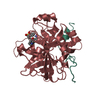
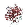
| Entry | Database: PDB / ID: 6sth | ||||||
|---|---|---|---|---|---|---|---|
| Title | Highly cationic RSL-R8 in complex with sulfonato-calix[8]arene | ||||||
 Components Components | Fucose-binding lectin protein | ||||||
 Keywords Keywords | SUGAR BINDING PROTEIN /  Molecular Glue / Calixarene / Supramolecular interactions / Sulfonato-calix[8]arene / ligand conformation Molecular Glue / Calixarene / Supramolecular interactions / Sulfonato-calix[8]arene / ligand conformation | ||||||
| Function / homology | Fucose-specific lectin / Fungal fucose-specific lectin / carbohydrate binding / sulfonato-calix[8]arene / Fucose-binding lectin protein Function and homology information Function and homology information | ||||||
| Biological species |  Ralstonia solanacearum (bacteria) Ralstonia solanacearum (bacteria) | ||||||
| Method | X-RAY DIFFRACTION / SYNCHROTRON / MOLECULAR REPLACEMENT / Resolution: 1.726 Å | ||||||
 Authors Authors | Skorek, T. / Engilberge, S. / Crowley, P.B. | ||||||
| Funding support |  Ireland, 1items Ireland, 1items
| ||||||
 Citation Citation | Journal: To Be Published Title: Highly cationic RSL-R8 in complex with sulfonato-calix[8]arene Authors: Skorek, T. / Engilberge, S. / Crowley, P.B. | ||||||
| History |
|
- Structure visualization
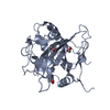
| Structure viewer | Molecule: MolmilJmol/JSmol |
|---|
- Downloads & links
-Download
| PDBx/mmCIF format | 6sth.cif.gz | 109.6 KB | Display | PDBx/mmCIF format |
|---|---|---|---|---|
| PDB format | pdb6sth.ent.gz | 85.2 KB | Display | PDB format |
| PDBx/mmJSON format | 6sth.json.gz | Tree view | PDBx/mmJSON format | |
| Others |  Other downloads Other downloads |
-Validation report
| Arichive directory | https://data.pdbj.org/pub/pdb/validation_reports/st/6sthftp://data.pdbj.org/pub/pdb/validation_reports/st/6sth | HTTPS FTP |
|---|
-Related structure data
| Similar structure data |
|---|


-Links
 PDBj
PDBj- Assembly
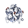
| Deposited unit | 
| |||||||||||||||
|---|---|---|---|---|---|---|---|---|---|---|---|---|---|---|---|---|
| 1 | 
| |||||||||||||||
| 2 | 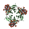
| |||||||||||||||
| Unit cell |
| |||||||||||||||
| Components on special symmetry positions |
|
-Components
| #1: Protein | Mass: 9887.790 Da / Num. of mol.: 2 Source method: isolated from a genetically manipulated source Source: (gene. exp.) Ralstonia solanacearum (bacteria)Gene: RSP795_21825, RSP799_05830, RSP822_19650, RUN39_v1_50103 Production host: Escherichia coli (E. coli) / References: UniProt: A0A0S4TLR1#2: Chemical | ChemComp-GOL / Glycerol  Mass: 92.094 Da / Num. of mol.: 6 / Source method: obtained synthetically / Formula: C3H8O3 Mass: 92.094 Da / Num. of mol.: 6 / Source method: obtained synthetically / Formula: C3H8O3#3: Chemical | Sulfate  Mass: 96.063 Da / Num. of mol.: 2 / Source method: obtained synthetically / Formula: SO4 Mass: 96.063 Da / Num. of mol.: 2 / Source method: obtained synthetically / Formula: SO4#4: Chemical | 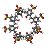  Mass: 1489.481 Da / Num. of mol.: 2 / Source method: obtained synthetically / Formula: C56H48O32S8 / Feature type: SUBJECT OF INVESTIGATION Mass: 1489.481 Da / Num. of mol.: 2 / Source method: obtained synthetically / Formula: C56H48O32S8 / Feature type: SUBJECT OF INVESTIGATION#5: Water | ChemComp-HOH / | Water Mass: 18.015 Da / Num. of mol.: 290 / Source method: isolated from a natural source / Formula: H2O Mass: 18.015 Da / Num. of mol.: 290 / Source method: isolated from a natural source / Formula: H2OHas ligand of interest | Y | |
|---|
-Experimental details
-Experiment
| Experiment | Method: X-RAY DIFFRACTION / Number of used crystals: 1 |
|---|
- Sample preparation
| Crystal | Density Matthews: 3.18 Å3/Da / Density % sol: 55 % |
|---|---|
| Crystal grow | Temperature: 293 K / Method: vapor diffusion, hanging drop Details: 0.6 mM RSL-R8 combined 33 mM sulfonato-calix[8]arene were crystallized using 0.1 M TRIS; pH 8.5, 1.26 M Ammonium sulfate, 0.2M Lithium sulfate. |
-Data collection
| Diffraction | Mean temperature: 100 K / Serial crystal experiment: N |
|---|---|
| Diffraction source | Source: SYNCHROTRON / Site: SOLEIL  / Beamline: PROXIMA 2 / Wavelength: 0.97624 Å / Beamline: PROXIMA 2 / Wavelength: 0.97624 Å |
| Detector | Type: DECTRIS EIGER X 9M / Detector: PIXEL / Date: Feb 2, 2019 |
| Radiation | Protocol: SINGLE WAVELENGTH / Monochromatic (M) / Laue (L): M / Scattering type: x-ray |
| Radiation wavelength | Wavelength: 0.97624 Å / Relative weight: 1 |
| Reflection | Resolution: 1.73→59.3 Å / Num. obs: 25591 / % possible obs: 99.9 % / Redundancy: 9.5 % / Biso Wilson estimate: 19.93 Å2 / Rpim(I) all: 0.062 / Rrim(I) all: 0.192 / Net I/σ(I): 13.7 |
| Reflection shell | Resolution: 1.73→1.76 Å / Num. unique obs: 1242 / Rpim(I) all: 0.055 / Rrim(I) all: 1.691 |
- Processing
| Software |
| ||||||||||||||||||||||||||||||||||||||||||||||||||||||||||||||||||||||
|---|---|---|---|---|---|---|---|---|---|---|---|---|---|---|---|---|---|---|---|---|---|---|---|---|---|---|---|---|---|---|---|---|---|---|---|---|---|---|---|---|---|---|---|---|---|---|---|---|---|---|---|---|---|---|---|---|---|---|---|---|---|---|---|---|---|---|---|---|---|---|---|
| Refinement | Method to determine structure: MOLECULAR REPLACEMENT / Resolution: 1.726→39.302 Å / SU ML: 0.24 / Cross valid method: THROUGHOUT / σ(F): 1.98 / Phase error: 29.28 / Stereochemistry target values: ML
| ||||||||||||||||||||||||||||||||||||||||||||||||||||||||||||||||||||||
| Solvent computation | Shrinkage radii: 0.9 Å / VDW probe radii: 1.11 Å / Solvent model: FLAT BULK SOLVENT MODEL | ||||||||||||||||||||||||||||||||||||||||||||||||||||||||||||||||||||||
| Refinement step | Cycle: LAST / Resolution: 1.726→39.302 Å
| ||||||||||||||||||||||||||||||||||||||||||||||||||||||||||||||||||||||
| Refine LS restraints |
| ||||||||||||||||||||||||||||||||||||||||||||||||||||||||||||||||||||||
| LS refinement shell |
| ||||||||||||||||||||||||||||||||||||||||||||||||||||||||||||||||||||||
| Refinement TLS params. | Method: refined / Origin x: 43.1291 Å / Origin y: 33.2013 Å / Origin z: 57.378 Å
| ||||||||||||||||||||||||||||||||||||||||||||||||||||||||||||||||||||||
| Refinement TLS group | Selection details: all |
