 Movie
Movie Controller


+ Open data

- Basic information
| Entry | Database: PDB / ID: 3gja | ||||||
|---|---|---|---|---|---|---|---|
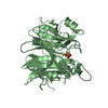
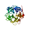
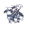
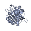
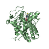
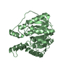
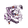
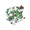
| Title | CytC3 | ||||||
 Components Components | CytC3 | ||||||
 Keywords Keywords |  BIOSYNTHETIC PROTEIN / CytC3 / halogenase / beta barrel BIOSYNTHETIC PROTEIN / CytC3 / halogenase / beta barrel | ||||||
| Function / homology |  Function and homology information Function and homology information | ||||||
| Biological species |  Streptomyces (bacteria) Streptomyces (bacteria) | ||||||
| Method | X-RAY DIFFRACTION / SYNCHROTRON / MOLECULAR REPLACEMENT / molecular replacement / Resolution: 2.2 Å | ||||||
 Authors Authors | Wong, C. / Drennan, C.L. | ||||||
 Citation Citation | Journal: J.Am.Chem.Soc. / Year: 2009 Title: Structural analysis of an open active site conformation of nonheme iron halogenase CytC3 Authors: Wong, C. / Fujimori, D.G. / Walsh, C.T. / Drennan, C.L. | ||||||
| History |
|
- Structure visualization
| Structure viewer | Molecule: MolmilJmol/JSmol |
|---|
- Downloads & links
-Download
| PDBx/mmCIF format | 3gja.cif.gz | 124.7 KB | Display | PDBx/mmCIF format |
|---|---|---|---|---|
| PDB format | pdb3gja.ent.gz | 95.7 KB | Display | PDB format |
| PDBx/mmJSON format | 3gja.json.gz | Tree view | PDBx/mmJSON format | |
| Others |  Other downloads Other downloads |
-Validation report
| Arichive directory | https://data.pdbj.org/pub/pdb/validation_reports/gj/3gjaftp://data.pdbj.org/pub/pdb/validation_reports/gj/3gja | HTTPS FTP |
|---|
-Related structure data
| Related structure data |  3gjbC 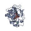 2fctS C: citing same article ( S: Starting model for refinement |
|---|---|
| Similar structure data |


-Links
 PDBj
PDBj- Assembly
| Deposited unit | 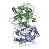
| ||||||||
|---|---|---|---|---|---|---|---|---|---|
| 1 | 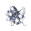
| ||||||||
| 2 | 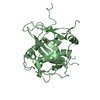
| ||||||||
| 3 | 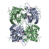
| ||||||||
| Unit cell |
|
-Components
| #1: Protein | Mass: 36484.574 Da / Num. of mol.: 2 Source method: isolated from a genetically manipulated source Source: (gene. exp.) Streptomyces (bacteria) / Strain: pSPHE02 / Gene: cytC3 / Plasmid: pET28b / Production host: Escherichia coli (E. coli) / Strain (production host): BL21(DE3) / References: UniProt: D0VX22*PLUS#2: Chemical | ChemComp-ACT / Acetate  Mass: 59.044 Da / Num. of mol.: 4 / Source method: obtained synthetically / Formula: C2H3O2 Mass: 59.044 Da / Num. of mol.: 4 / Source method: obtained synthetically / Formula: C2H3O2#3: Water | ChemComp-HOH / | Water Mass: 18.015 Da / Num. of mol.: 230 / Source method: isolated from a natural source / Formula: H2O Mass: 18.015 Da / Num. of mol.: 230 / Source method: isolated from a natural source / Formula: H2O |
|---|
-Experimental details
-Experiment
| Experiment | Method: X-RAY DIFFRACTION / Number of used crystals: 1 |
|---|
- Sample preparation
| Crystal | Density Matthews: 3.46 Å3/Da / Density % sol: 64.45 % |
|---|---|
| Crystal grow | Temperature: 298 K / Method: vapor diffusion, hanging drop / pH: 7 Details: 2.8 M sodium acetate trihydrate, pH 7.0, VAPOR DIFFUSION, HANGING DROP, temperature 298K |
-Data collection
| Diffraction | Mean temperature: 100 K | ||||||||||||||||||||||||||||||||||||||||||||||||||||||||||||||||||
|---|---|---|---|---|---|---|---|---|---|---|---|---|---|---|---|---|---|---|---|---|---|---|---|---|---|---|---|---|---|---|---|---|---|---|---|---|---|---|---|---|---|---|---|---|---|---|---|---|---|---|---|---|---|---|---|---|---|---|---|---|---|---|---|---|---|---|---|
| Diffraction source | Source: SYNCHROTRON / Site: SSRL  / Beamline: BL11-1 / Wavelength: 1 Å / Beamline: BL11-1 / Wavelength: 1 Å | ||||||||||||||||||||||||||||||||||||||||||||||||||||||||||||||||||
| Detector | Type: MARMOSAIC 325 mm CCD / Detector: CCD / Date: Jun 1, 2006 / Details: inter-image dead time 5s | ||||||||||||||||||||||||||||||||||||||||||||||||||||||||||||||||||
| Radiation | Monochromator: side scattering bent cube-root I-beam single crystal Protocol: SINGLE WAVELENGTH / Scattering type: x-ray | ||||||||||||||||||||||||||||||||||||||||||||||||||||||||||||||||||
| Radiation wavelength | Wavelength: 1 Å / Relative weight: 1 | ||||||||||||||||||||||||||||||||||||||||||||||||||||||||||||||||||
| Reflection | Resolution: 2.2→50 Å / Num. obs: 50903 / % possible obs: 95.9 % / Rmerge(I) obs: 0.056 / Χ2: 1.136 | ||||||||||||||||||||||||||||||||||||||||||||||||||||||||||||||||||
| Reflection shell |
|
-Phasing
| Phasing | Method: molecular replacement | |||||||||
|---|---|---|---|---|---|---|---|---|---|---|
| Phasing MR | Model details: Phaser MODE: MR_AUTO
|
- Processing
| Software |
| ||||||||||||||||||||||||
|---|---|---|---|---|---|---|---|---|---|---|---|---|---|---|---|---|---|---|---|---|---|---|---|---|---|
| Refinement | Method to determine structure: MOLECULAR REPLACEMENT Starting model: PDB entry 2FCT Resolution: 2.2→44.52 Å / Occupancy max: 1 / Occupancy min: 1 / FOM work R set: 0.806 / σ(F): 0
| ||||||||||||||||||||||||
| Solvent computation | Bsol: 50.807 Å2 | ||||||||||||||||||||||||
| Displacement parameters | Biso max: 96.75 Å2 / Biso mean: 45.732 Å2 / Biso min: 25.51 Å2
| ||||||||||||||||||||||||
| Refine analyze |
| ||||||||||||||||||||||||
| Refinement step | Cycle: LAST / Resolution: 2.2→44.52 Å
| ||||||||||||||||||||||||
| Refine LS restraints |
| ||||||||||||||||||||||||
| Xplor file |
|
