 Movie
Movie Controller

[English] 日本語
 Yorodumi
- PDB-4kcb: Crystal Structure of Exo-1,5-alpha-L-arabinanase from Bovine Rumi... -
+ Open data

- Basic information
| Entry | Database: PDB / ID: 4kcb | ||||||
|---|---|---|---|---|---|---|---|
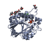
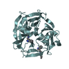
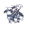
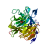
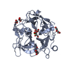
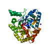
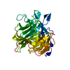
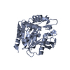
| Title | Crystal Structure of Exo-1,5-alpha-L-arabinanase from Bovine Ruminal Metagenomic Library | ||||||
 Components Components | Arabinan endo-1,5-alpha-L-arabinosidase | ||||||
 Keywords Keywords | HYDROLASE / beta-propeller / GH43 / glycoside hydrolase / arabinanase | ||||||
| Function / homology |  Function and homology informationarabinan endo-1,5-alpha-L-arabinosidase activity / arabinan catabolic process Function and homology informationarabinan endo-1,5-alpha-L-arabinosidase activity / arabinan catabolic processSimilarity search - Function | ||||||
| Biological species |  uncultured bacterium (environmental samples) uncultured bacterium (environmental samples) | ||||||
| Method | X-RAY DIFFRACTION / SYNCHROTRON / MOLECULAR REPLACEMENT / Resolution: 2.9 Å | ||||||
 Authors Authors | Santos, C.R. / Polo, C.C. / Costa, M.C.M.F. / Nascimento, A.F.Z. / Wong, D.W.S. / Murakami, M.T. | ||||||
 Citation Citation | Journal: J.Biol.Chem. / Year: 2014 Title: Mechanistic strategies for catalysis adopted by evolutionary distinct family 43 arabinanases. Authors: Santos, C.R. / Polo, C.C. / Costa, M.C. / Nascimento, A.F. / Meza, A.N. / Cota, J. / Hoffmam, Z.B. / Honorato, R.V. / Oliveira, P.S. / Goldman, G.H. / Gilbert, H.J. / Prade, R.A. / Ruller, R. ...Authors: Santos, C.R. / Polo, C.C. / Costa, M.C. / Nascimento, A.F. / Meza, A.N. / Cota, J. / Hoffmam, Z.B. / Honorato, R.V. / Oliveira, P.S. / Goldman, G.H. / Gilbert, H.J. / Prade, R.A. / Ruller, R. / Squina, F.M. / Wong, D.W. / Murakami, M.T. | ||||||
| History |
|
- Structure visualization
| Structure viewer | Molecule: MolmilJmol/JSmol |
|---|
- Downloads & links
-Download
| PDBx/mmCIF format | 4kcb.cif.gz | 132.2 KB | Display | PDBx/mmCIF format |
|---|---|---|---|---|
| PDB format | pdb4kcb.ent.gz | 106.9 KB | Display | PDB format |
| PDBx/mmJSON format | 4kcb.json.gz | Tree view | PDBx/mmJSON format | |
| Others |  Other downloads Other downloads |
-Validation report
| Arichive directory | https://data.pdbj.org/pub/pdb/validation_reports/kc/4kcbftp://data.pdbj.org/pub/pdb/validation_reports/kc/4kcb | HTTPS FTP |
|---|
-Related structure data


-Links
 PDBj
PDBj- Assembly
| Deposited unit | 
| ||||||||
|---|---|---|---|---|---|---|---|---|---|
| 1 | 
| ||||||||
| 2 | 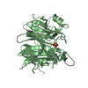
| ||||||||
| Unit cell |
| ||||||||
| Details | the biological unit is a monomer; there are 2 biological units in asymmetric unit |
-Components
| #1: Protein | Mass: 50320.492 Da / Num. of mol.: 2 Source method: isolated from a genetically manipulated source Source: (gene. exp.) uncultured bacterium (environmental samples)Description: THE SAMPLE SEQUENCE WAS IDENTIFIED FROM A DNA LIBRARY CONSTRUCTED FROM BOVINE RUMEN FLUID (WONG ET AL., 2008) Production host: Escherichia coli (E. coli)References: UniProt: D2XML8, arabinan endo-1,5-alpha-L-arabinanase #2: Chemical | Phosphate  Mass: 94.971 Da / Num. of mol.: 3 / Source method: obtained synthetically / Formula: PO4 Mass: 94.971 Da / Num. of mol.: 3 / Source method: obtained synthetically / Formula: PO4#3: Water | ChemComp-HOH / | Water Mass: 18.015 Da / Num. of mol.: 19 / Source method: isolated from a natural source / Formula: H2O Mass: 18.015 Da / Num. of mol.: 19 / Source method: isolated from a natural source / Formula: H2O |
|---|
-Experimental details
-Experiment
| Experiment | Method: X-RAY DIFFRACTION / Number of used crystals: 1 |
|---|
- Sample preparation
| Crystal | Density Matthews: 2.24 Å3/Da / Density % sol: 45.05 % |
|---|---|
| Crystal grow | Temperature: 291 K / Method: vapor diffusion, sitting drop / pH: 8.5 Details: Ammonium Phosphate, pH 8.5, VAPOR DIFFUSION, SITTING DROP, temperature 291K |
-Data collection
| Diffraction | Mean temperature: 100 K |
|---|---|
| Diffraction source | Source: SYNCHROTRON / Site: LNLS  / Beamline: W01B-MX2 / Wavelength: 1.459 Å / Beamline: W01B-MX2 / Wavelength: 1.459 Å |
| Detector | Type: MARMOSAIC 225 mm CCD / Detector: CCD / Date: Jan 1, 2013 |
| Radiation | Monochromator: Si(111)double-crystal / Protocol: SINGLE WAVELENGTH / Monochromatic (M) / Laue (L): M / Scattering type: x-ray |
| Radiation wavelength | Wavelength: 1.459 Å / Relative weight: 1 |
| Reflection | Resolution: 2.9→40 Å / Num. obs: 20619 / % possible obs: 97.5 % / Observed criterion σ(F): 0 / Observed criterion σ(I): 0 |
| Reflection shell | Resolution: 2.9→3 Å / % possible all: 99.2 |
- Processing
| Software |
| |||||||||||||||||||||||||||||||||||||||||||||||||||||||||||||||||
|---|---|---|---|---|---|---|---|---|---|---|---|---|---|---|---|---|---|---|---|---|---|---|---|---|---|---|---|---|---|---|---|---|---|---|---|---|---|---|---|---|---|---|---|---|---|---|---|---|---|---|---|---|---|---|---|---|---|---|---|---|---|---|---|---|---|---|
| Refinement | Method to determine structure: MOLECULAR REPLACEMENT / Resolution: 2.9→35.03 Å / Cor.coef. Fo:Fc: 0.922 / Cor.coef. Fo:Fc free: 0.885 / SU B: 15.203 / SU ML: 0.29 / Cross valid method: THROUGHOUT / σ(F): 0 / ESU R Free: 0.396 / Stereochemistry target values: MAXIMUM LIKELIHOOD
| |||||||||||||||||||||||||||||||||||||||||||||||||||||||||||||||||
| Solvent computation | Ion probe radii: 0.8 Å / Shrinkage radii: 0.8 Å / VDW probe radii: 1.4 Å / Solvent model: MASK | |||||||||||||||||||||||||||||||||||||||||||||||||||||||||||||||||
| Displacement parameters | Biso mean: 54.944 Å2
| |||||||||||||||||||||||||||||||||||||||||||||||||||||||||||||||||
| Refinement step | Cycle: LAST / Resolution: 2.9→35.03 Å
| |||||||||||||||||||||||||||||||||||||||||||||||||||||||||||||||||
| Refine LS restraints |
| |||||||||||||||||||||||||||||||||||||||||||||||||||||||||||||||||
| LS refinement shell | Resolution: 2.896→2.97 Å / Total num. of bins used: 20
|
