National Institutes of Health/National Institute of General Medical Sciences (NIH/NIGMS)
R01 GM067808
United States
National Institutes of Health/National Institute Of Allergy and Infectious Diseases (NIH/NIAID)
AI078000
United States
UC Irvine Center for Biomembrane Systems
United States
Citation
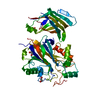
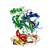
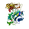
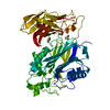
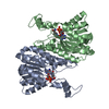
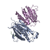
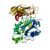
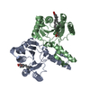
Journal: To Be Published Title: Mechanistic insights from the crystal structure of an inward proton-transportingAnabaena sensory rhodopsin mutant Authors: Dong, B.H. / Luecke, H.
Resolution: 2.3→33.19 Å / Cor.coef. Fo:Fc: 0.943 / Cor.coef. Fo:Fc free: 0.924 / SU B: 6.228 / SU ML: 0.155 / Cross valid method: THROUGHOUT / ESU R: 0.329 / ESU R Free: 0.232 / Stereochemistry target values: MAXIMUM LIKELIHOOD / Details: HYDROGENS HAVE BEEN USED IF PRESENT IN THE INPUT
Rfactor
Num. reflection
% reflection
Selection details
Rfree
0.24951
1449
5.1 %
RANDOM
Rwork
0.21976
-
-
-
obs
0.22137
26889
96.59 %
-
Solvent computation
Ion probe radii: 0.8 Å / Shrinkage radii: 0.8 Å / VDW probe radii: 1.2 Å / Solvent model: MASK
 Movie
Movie Controller

 Yorodumi
 Open data
 Basic information
 Components
 Keywords
 SIGNALING PROTEIN /
  Function and homology information
 Nostoc sp. (bacteria)
 Authors
 United States, 3items
  Citation
 Structure visualization
 Downloads & links
 Other downloads

 PDBj
PDBj


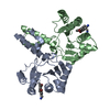
 Assembly

 Mass: 284.436 Da / Num. of mol.: 2 / Source method: obtained synthetically / Formula: C20H28O
 Mass: 744.034 Da / Num. of mol.: 35 / Source method: obtained synthetically / Formula: C41H78NO8P / Comment: DOPE, phospholipid*YM
 Mass: 18.015 Da / Num. of mol.: 109 / Source method: isolated from a natural source / Formula: H2O
 Sample preparation
 Processing