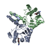
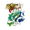
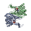
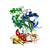
[English] 日本語
Yorodumi
- PDB-5okp: Crystal structure of human SHIP2 Phosphatase-C2 double mutant F59... -

+
Open data


ID or keywords:

Loading...

-
Basic information

Entry
Database: PDB / ID: 5okp
TitleCrystal structure of human SHIP2 Phosphatase-C2 double mutant F593D/L597D
ComponentsPhosphatidylinositol 3,4,5-trisphosphate 5-phosphatase 2
KeywordsHYDROLASE / SHIP2 / Phosphatase / C2 / phosphatidylinositol (3 / 4 / 5)-triphosphate
Function / homology
Function and homology information


negative regulation of insulin-like growth factor receptor signaling pathway / phosphatidylinositol-3,4,5-trisphosphate 5-phosphatase / inositol-polyphosphate 5-phosphatase activity / ruffle assembly / phosphatidylinositol-3,4,5-trisphosphate 5-phosphatase activity / regulation of actin filament organization / phosphatidylinositol dephosphorylation / endochondral ossification / phosphatidylinositol biosynthetic process / immune system process ...negative regulation of insulin-like growth factor receptor signaling pathway / phosphatidylinositol-3,4,5-trisphosphate 5-phosphatase / inositol-polyphosphate 5-phosphatase activity / ruffle assembly / phosphatidylinositol-3,4,5-trisphosphate 5-phosphatase activity / regulation of actin filament organization / phosphatidylinositol dephosphorylation / endochondral ossification / phosphatidylinositol biosynthetic process / immune system process / Synthesis of IP3 and IP4 in the cytosol / establishment of mitotic spindle orientation / Synthesis of PIPs at the plasma membrane / regulation of immune response / Interleukin receptor SHC signaling / ERK1 and ERK2 cascade / SH2 domain binding / basal plasma membrane / post-embryonic development / phosphatidylinositol 3-kinase/protein kinase B signal transduction / filopodium / actin filament organization / response to insulin / SH3 domain binding / spindle pole / endocytosis / glucose metabolic process / Signaling by CSF1 (M-CSF) in myeloid cells / regulation of protein localization / lamellipodium / actin binding / gene expression / cell adhesion / nuclear speck / negative regulation of cell population proliferation / negative regulation of gene expression / apoptotic process / Golgi apparatus / nucleus / cytosol
Similarity search - Function
Inositol polyphosphate-related phosphatase / Inositol polyphosphate phosphatase, catalytic domain homologues / Deoxyribonuclease I; Chain A / Endonuclease/exonuclease/phosphatase / Endonuclease/Exonuclease/phosphatase family / SAM domain (Sterile alpha motif) / Endonuclease/exonuclease/phosphatase superfamily / SAM domain profile. / Sterile alpha motif. / Sterile alpha motif domain ...Inositol polyphosphate-related phosphatase / Inositol polyphosphate phosphatase, catalytic domain homologues / Deoxyribonuclease I; Chain A / Endonuclease/exonuclease/phosphatase / Endonuclease/Exonuclease/phosphatase family / SAM domain (Sterile alpha motif) / Endonuclease/exonuclease/phosphatase superfamily / SAM domain profile. / Sterile alpha motif. / Sterile alpha motif domain / Sterile alpha motif/pointed domain superfamily / SH2 domain / Src homology 2 (SH2) domain profile. / Src homology 2 domains / SH2 domain / SH2 domain superfamily / 4-Layer Sandwich / Alpha Beta
Similarity search - Domain/homology
Phosphatidylinositol 3,4,5-trisphosphate 5-phosphatase 2
Similarity search - Component
Biological speciesHomo sapiens (human)
MethodX-RAY DIFFRACTION / SYNCHROTRON / MOLECULAR REPLACEMENT / Resolution: 1.85 Å
AuthorsLe Coq, J. / Lietha, D.
CitationJournal: Elife / Year: 2017
Title: Structural basis for interdomain communication in SHIP2 providing high phosphatase activity.
Authors: Le Coq, J. / Camacho-Artacho, M. / Velazquez, J.V. / Santiveri, C.M. / Gallego, L.H. / Campos-Olivas, R. / Dolker, N. / Lietha, D.
History
DepositionJul 25, 2017Deposition site: PDBE / Processing site: PDBE
Revision 1.0Aug 23, 2017Provider: repository / Type: Initial release
Revision 1.1May 8, 2024Group: Data collection / Database references / Category: chem_comp_atom / chem_comp_bond / database_2
Item: _database_2.pdbx_DOI / _database_2.pdbx_database_accession

-
Structure visualization

Structure viewerMolecule:
MolmilJmol/JSmol

Downloads & links

-
Assembly

Deposited unit
A: Phosphatidylinositol 3,4,5-trisphosphate 5-phosphatase 2
hetero molecules


Theoretical massNumber of molelcules
Total (without water)52,9695
Polymers52,7201
Non-polymers2484
Water2,504139
1


  • Idetical with deposited unit
  • defined by author&software
  • Evidence: gel filtration
TypeNameSymmetry operationNumber
identity operation1_555x,y,z1
Buried area710 Å2
ΔGint10 kcal/mol
Surface area18860 Å2
MethodPISA
Unit cell
Length a, b, c (Å)43.745, 73.426, 157.964
Angle α, β, γ (deg.)90.00, 90.70, 90.00
Int Tables number5
Space group name H-MI121
Components on special symmetry positions
IDModelComponents
11A-1123-

HOH

21A-1136-

HOH

-
Components

#1: Protein Phosphatidylinositol 3,4,5-trisphosphate 5-phosphatase 2 / Inositol polyphosphate phosphatase-like protein 1 / INPPL-1 / Protein 51C / SH2 domain-containing ...Inositol polyphosphate phosphatase-like protein 1 / INPPL-1 / Protein 51C / SH2 domain-containing inositol 5'-phosphatase 2 / SHIP-2


Mass: 52720.344 Da / Num. of mol.: 1 / Mutation: F593D, L597D
Source method: isolated from a genetically manipulated source
Details: The first two residues are from the expression vector in which the gene was cloned.
Source: (gene. exp.) Homo sapiens (human) / Gene: INPPL1, SHIP2 / Plasmid: pOPINJ / Production host: Escherichia coli BL21(DE3) (bacteria) / Variant (production host): Rosetta2(DE3)pLysS
References: UniProt: O15357, phosphatidylinositol-3,4,5-trisphosphate 5-phosphatase
#2: Chemical
ChemComp-EDO / 1,2-ETHANEDIOL / ETHYLENE GLYCOL / Ethylene glycol


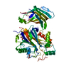
Mass: 62.068 Da / Num. of mol.: 4 / Source method: obtained synthetically / Formula: C2H6O2
#3: Water ChemComp-HOH / water / Water


Mass: 18.015 Da / Num. of mol.: 139 / Source method: isolated from a natural source / Formula: H2O

-
Experimental details

-
Experiment

ExperimentMethod: X-RAY DIFFRACTION / Number of used crystals: 1

-
Sample preparation

CrystalDensity Matthews: 2.42 Å3/Da / Density % sol: 49.25 %
Crystal growTemperature: 293 K / Method: vapor diffusion, sitting drop / pH: 7.1 / Details: 50 mM HEPES pH 7.1, 17.5% PEG 1000

-
Data collection

DiffractionMean temperature: 100 K
Diffraction sourceSource: SYNCHROTRON / Site: ESRF / Beamline: ID29 / Wavelength: 0.976 Å
DetectorType: DECTRIS PILATUS 6M / Detector: PIXEL / Date: Jul 28, 2015
RadiationProtocol: SINGLE WAVELENGTH / Monochromatic (M) / Laue (L): M / Scattering type: x-ray
Radiation wavelengthWavelength: 0.976 Å / Relative weight: 1
ReflectionResolution: 1.85→78.98 Å / Num. obs: 40283 / % possible obs: 99.3 % / Redundancy: 3.4 % / CC1/2: 0.999 / Rmerge(I) obs: 0.059 / Rpim(I) all: 0.038 / Net I/σ(I): 11.6
Reflection shellResolution: 1.85→1.89 Å / Redundancy: 3.5 % / Rmerge(I) obs: 0.595 / Mean I/σ(I) obs: 2.1 / CC1/2: 0.768 / Rpim(I) all: 0.372 / % possible all: 99.1

-
Processing

Software
NameVersionClassification
REFMAC5.8.0158refinement
XDSdata reduction
Aimlessdata scaling
PHASERphasing
RefinementMethod to determine structure: MOLECULAR REPLACEMENT / Resolution: 1.85→78.98 Å / Cor.coef. Fo:Fc: 0.961 / Cor.coef. Fo:Fc free: 0.946 / SU B: 5.945 / SU ML: 0.085 / Cross valid method: THROUGHOUT / ESU R: 0.125 / ESU R Free: 0.115 / Details: HYDROGENS HAVE BEEN ADDED IN THE RIDING POSITIONS
RfactorNum. reflection% reflectionSelection details
Rfree0.20469 2118 5 %RANDOM
Rwork0.17787 ---
obs0.17923 40283 99.14 %-
Solvent computationIon probe radii: 0.8 Å / Shrinkage radii: 0.8 Å / VDW probe radii: 1.2 Å
Displacement parametersBiso mean: 33.328 Å2
Baniso -1Baniso -2Baniso -3
1--0.21 Å20 Å2-0.11 Å2
2---0.68 Å20 Å2
3---0.89 Å2
Refinement stepCycle: 1 / Resolution: 1.85→78.98 Å
ProteinNucleic acidLigandSolventTotal
Num. atoms3394 0 16 139 3549
Refine LS restraints
Refine-IDTypeDev idealDev ideal targetNumber
X-RAY DIFFRACTIONr_bond_refined_d0.0080.0193516
X-RAY DIFFRACTIONr_bond_other_d0.0030.023218
X-RAY DIFFRACTIONr_angle_refined_deg1.261.9494757
X-RAY DIFFRACTIONr_angle_other_deg0.88337473
X-RAY DIFFRACTIONr_dihedral_angle_1_deg6.0575426
X-RAY DIFFRACTIONr_dihedral_angle_2_deg33.73624.012167
X-RAY DIFFRACTIONr_dihedral_angle_3_deg13.21815610
X-RAY DIFFRACTIONr_dihedral_angle_4_deg18.7711521
X-RAY DIFFRACTIONr_chiral_restr0.0730.2529
X-RAY DIFFRACTIONr_gen_planes_refined0.0050.023855
X-RAY DIFFRACTIONr_gen_planes_other0.0010.02742
X-RAY DIFFRACTIONr_nbd_refined
X-RAY DIFFRACTIONr_nbd_other
X-RAY DIFFRACTIONr_nbtor_refined
X-RAY DIFFRACTIONr_nbtor_other
X-RAY DIFFRACTIONr_xyhbond_nbd_refined
X-RAY DIFFRACTIONr_xyhbond_nbd_other
X-RAY DIFFRACTIONr_metal_ion_refined
X-RAY DIFFRACTIONr_metal_ion_other
X-RAY DIFFRACTIONr_symmetry_vdw_refined
X-RAY DIFFRACTIONr_symmetry_vdw_other
X-RAY DIFFRACTIONr_symmetry_hbond_refined
X-RAY DIFFRACTIONr_symmetry_hbond_other
X-RAY DIFFRACTIONr_symmetry_metal_ion_refined
X-RAY DIFFRACTIONr_symmetry_metal_ion_other
X-RAY DIFFRACTIONr_mcbond_it0.8682.0511692
X-RAY DIFFRACTIONr_mcbond_other0.8682.051691
X-RAY DIFFRACTIONr_mcangle_it1.5243.0582107
X-RAY DIFFRACTIONr_mcangle_other1.5233.0592108
X-RAY DIFFRACTIONr_scbond_it0.9732.2221823
X-RAY DIFFRACTIONr_scbond_other0.9722.2221823
X-RAY DIFFRACTIONr_scangle_it
X-RAY DIFFRACTIONr_scangle_other1.6233.2672646
X-RAY DIFFRACTIONr_long_range_B_refined3.81823.3543747
X-RAY DIFFRACTIONr_long_range_B_other3.75323.1593728
X-RAY DIFFRACTIONr_rigid_bond_restr
X-RAY DIFFRACTIONr_sphericity_free
X-RAY DIFFRACTIONr_sphericity_bonded
LS refinement shellResolution: 1.85→1.898 Å / Total num. of bins used: 20
RfactorNum. reflection% reflection
Rfree0.24 147 -
Rwork0.24 2993 -
obs--99.02 %
Refinement TLS params.

Method: refined / Refine-ID: X-RAY DIFFRACTION

IDL112)L122)L132)L222)L232)L332)S11 (Å °)S12 (Å °)S13 (Å °)S21 (Å °)S22 (Å °)S23 (Å °)S31 (Å °)S32 (Å °)S33 (Å °)T112)T122)T132)T222)T232)T332)Origin x (Å)Origin y (Å)Origin z (Å)
10.81940.0156-0.00110.96050.09830.35-0.02210.0053-0.00390.00450.01090.05920.0204-0.02460.01120.00450.00420.01770.0657-0.00260.13710.113316.389641.3534
23.28381.6273-0.30225.61440.15762.39610.0141-0.08030.13730.7980.1392-0.016-0.0532-0.041-0.15330.1610.0470.02220.12930.00320.0492-22.782513.805965.9312
Refinement TLS group
IDRefine-IDRefine TLS-IDAuth asym-IDAuth seq-ID
1X-RAY DIFFRACTION1A419 - 731
2X-RAY DIFFRACTION2A746 - 874

+
About Yorodumi

-
News

-
Feb 9, 2022. New format data for meta-information of EMDB entries

New format data for meta-information of EMDB entries

  • Version 3 of the EMDB header file is now the official format.
  • The previous official version 1.9 will be removed from the archive.

Related info.:EMDB header

External links:wwPDB to switch to version 3 of the EMDB data model

-
Aug 12, 2020. Covid-19 info

Covid-19 info

URL: https://pdbj.org/emnavi/covid19.php

New page: Covid-19 featured information page in EM Navigator.

Related info.:Covid-19 info / Mar 5, 2020. Novel coronavirus structure data

+
Mar 5, 2020. Novel coronavirus structure data

Novel coronavirus structure data

Related info.:Yorodumi Speices / Aug 12, 2020. Covid-19 info

External links:COVID-19 featured content - PDBj / Molecule of the Month (242):Coronavirus Proteases

+
Jan 31, 2019. EMDB accession codes are about to change! (news from PDBe EMDB page)

EMDB accession codes are about to change! (news from PDBe EMDB page)

  • The allocation of 4 digits for EMDB accession codes will soon come to an end. Whilst these codes will remain in use, new EMDB accession codes will include an additional digit and will expand incrementally as the available range of codes is exhausted. The current 4-digit format prefixed with “EMD-” (i.e. EMD-XXXX) will advance to a 5-digit format (i.e. EMD-XXXXX), and so on. It is currently estimated that the 4-digit codes will be depleted around Spring 2019, at which point the 5-digit format will come into force.
  • The EM Navigator/Yorodumi systems omit the EMD- prefix.

Related info.:Q: What is EMD? / ID/Accession-code notation in Yorodumi/EM Navigator

External links:EMDB Accession Codes are Changing Soon! / Contact to PDBj

+
Jul 12, 2017. Major update of PDB

Major update of PDB

  • wwPDB released updated PDB data conforming to the new PDBx/mmCIF dictionary.
  • This is a major update changing the version number from 4 to 5, and with Remediation, in which all the entries are updated.
  • In this update, many items about electron microscopy experimental information are reorganized (e.g. em_software).
  • Now, EM Navigator and Yorodumi are based on the updated data.

External links:wwPDB Remediation / Enriched Model Files Conforming to OneDep Data Standards Now Available in the PDB FTP Archive

-
Yorodumi

Thousand views of thousand structures

  • Yorodumi is a browser for structure data from EMDB, PDB, SASBDB, etc.
  • This page is also the successor to EM Navigator detail page, and also detail information page/front-end page for Omokage search.
  • The word "yorodu" (or yorozu) is an old Japanese word meaning "ten thousand". "mi" (miru) is to see.

Related info.:EMDB / PDB / SASBDB / Comparison of 3 databanks / Yorodumi Search / Aug 31, 2016. New EM Navigator & Yorodumi / Yorodumi Papers / Jmol/JSmol / Function and homology information / Changes in new EM Navigator and Yorodumi

Read more