[English] 日本語
Yorodumi
- PDB-4qm9: Crystal Structure of a Putative Cysteine Dioxygenase From Bacillu... -

+
Open data


ID or keywords:

Loading...

-
Basic information

Entry
Database: PDB / ID: 4qm9
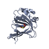
Title
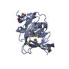
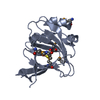
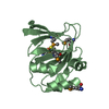
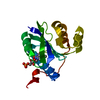
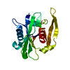
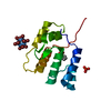
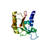
Crystal Structure of a Putative Cysteine Dioxygenase From Bacillus subtilis with Cys-bound
ComponentsCysteine dioxygenase
KeywordsOXIDOREDUCTASE / Cupin fold / catalyzes oxidation of cysteine to cysteine sulfinate
Function / homology
Function and homology information


cysteine dioxygenase / cysteine dioxygenase activity / L-cysteine catabolic process / oxidoreductase activity, acting on single donors with incorporation of molecular oxygen, incorporation of two atoms of oxygen / ferrous iron binding
Similarity search - Function
Cysteine dioxygenase type I / Cysteine dioxygenase type I / RmlC-like cupin domain superfamily / Jelly Rolls / RmlC-like jelly roll fold / Jelly Rolls / Sandwich / Mainly Beta
Similarity search - Domain/homology
CYSTEINE / : / Cysteine dioxygenase
Similarity search - Component
Biological speciesBacillus subtilis subsp. subtilis (bacteria)
MethodX-RAY DIFFRACTION / SYNCHROTRON / MOLECULAR REPLACEMENT / Resolution: 2.3 Å
AuthorsHartman, S.H. / Driggers, C.M. / Karplus, P.A.
CitationJournal: Protein Sci. / Year: 2015
Title: Structures of Arg- and Gln-type bacterial cysteine dioxygenase homologs.
Authors: Driggers, C.M. / Hartman, S.J. / Karplus, P.A.
History
DepositionJun 15, 2014Deposition site: RCSB / Processing site: RCSB
Revision 1.0Nov 19, 2014Provider: repository / Type: Initial release
Revision 1.1Dec 31, 2014Group: Database references
Revision 1.2Jan 14, 2015Group: Database references
Revision 1.3Mar 4, 2020Group: Database references / Structure summary / Category: struct / struct_ref_seq_dif / Item: _struct.title / _struct_ref_seq_dif.details
Revision 1.4Sep 20, 2023Group: Data collection / Database references ...Data collection / Database references / Derived calculations / Refinement description
Category: chem_comp_atom / chem_comp_bond ...chem_comp_atom / chem_comp_bond / database_2 / pdbx_initial_refinement_model / pdbx_struct_conn_angle / struct_conn / struct_site
Item: _database_2.pdbx_DOI / _database_2.pdbx_database_accession ..._database_2.pdbx_DOI / _database_2.pdbx_database_accession / _pdbx_struct_conn_angle.ptnr1_auth_comp_id / _pdbx_struct_conn_angle.ptnr1_auth_seq_id / _pdbx_struct_conn_angle.ptnr1_label_asym_id / _pdbx_struct_conn_angle.ptnr1_label_atom_id / _pdbx_struct_conn_angle.ptnr1_label_comp_id / _pdbx_struct_conn_angle.ptnr1_label_seq_id / _pdbx_struct_conn_angle.ptnr3_auth_comp_id / _pdbx_struct_conn_angle.ptnr3_auth_seq_id / _pdbx_struct_conn_angle.ptnr3_label_asym_id / _pdbx_struct_conn_angle.ptnr3_label_atom_id / _pdbx_struct_conn_angle.ptnr3_label_comp_id / _pdbx_struct_conn_angle.ptnr3_label_seq_id / _pdbx_struct_conn_angle.value / _struct_conn.pdbx_dist_value / _struct_conn.ptnr1_auth_asym_id / _struct_conn.ptnr1_auth_comp_id / _struct_conn.ptnr1_auth_seq_id / _struct_conn.ptnr1_label_asym_id / _struct_conn.ptnr1_label_atom_id / _struct_conn.ptnr1_label_comp_id / _struct_conn.ptnr1_label_seq_id / _struct_conn.ptnr2_auth_asym_id / _struct_conn.ptnr2_auth_comp_id / _struct_conn.ptnr2_auth_seq_id / _struct_conn.ptnr2_label_asym_id / _struct_conn.ptnr2_label_atom_id / _struct_conn.ptnr2_label_comp_id / _struct_site.pdbx_auth_asym_id / _struct_site.pdbx_auth_comp_id / _struct_site.pdbx_auth_seq_id

-
Structure visualization

Structure viewerMolecule:
MolmilJmol/JSmol

Downloads & links

-
Assembly

Deposited unit
A: Cysteine dioxygenase
B: Cysteine dioxygenase
hetero molecules


Theoretical massNumber of molelcules
Total (without water)39,5826
Polymers39,2282
Non-polymers3544
Water1,20767
1
A: Cysteine dioxygenase
hetero molecules


Theoretical massNumber of molelcules
Total (without water)19,7913
Polymers19,6141
Non-polymers1772
Water181
TypeNameSymmetry operationNumber
identity operation1_555x,y,z1
MethodPISA
2
B: Cysteine dioxygenase
hetero molecules


Theoretical massNumber of molelcules
Total (without water)19,7913
Polymers19,6141
Non-polymers1772
Water181
TypeNameSymmetry operationNumber
identity operation1_555x,y,z1
MethodPISA
Unit cell
Length a, b, c (Å)65.530, 65.530, 199.360
Angle α, β, γ (deg.)90.00, 90.00, 90.00
Int Tables number95
Space group name H-MP4322

-
Components

#1: Protein Cysteine dioxygenase / / CDO


Mass: 19614.113 Da / Num. of mol.: 2
Source method: isolated from a genetically manipulated source
Source: (gene. exp.) Bacillus subtilis subsp. subtilis (bacteria)
Strain: 168 / Gene: BSU31140, cdoA, Cysteine Dioxygenase, yubC / Plasmid: pET32a / Production host: Escherichia coli (E. coli) / Strain (production host): Bl21-DE3 / References: UniProt: O32085, cysteine dioxygenase
#2: Chemical ChemComp-FE / FE (III) ION / Iron


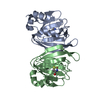
Mass: 55.845 Da / Num. of mol.: 2 / Source method: obtained synthetically / Formula: Fe
#3: Chemical ChemComp-CYS / CYSTEINE / Cysteine


Type: L-peptide linking / Mass: 121.158 Da / Num. of mol.: 2 / Source method: obtained synthetically / Formula: C3H7NO2S
#4: Water ChemComp-HOH / water / Water


Mass: 18.015 Da / Num. of mol.: 67 / Source method: isolated from a natural source / Formula: H2O

-
Experimental details

-
Experiment

ExperimentMethod: X-RAY DIFFRACTION / Number of used crystals: 1

-
Sample preparation

CrystalDensity Matthews: 2.73 Å3/Da / Density % sol: 54.91 %
Crystal growTemperature: 298 K / Method: vapor diffusion, hanging drop / pH: 6
Details: Purified enzyme was concentrated to 10 mg/mL and 4 uL was mixed with 4 uL of a reservoir containing 18% (w/v) PEG 4000, 0.1 M potassium acetate, 0.05 M MES, pH 6.0, VAPOR DIFFUSION, HANGING ...Details: Purified enzyme was concentrated to 10 mg/mL and 4 uL was mixed with 4 uL of a reservoir containing 18% (w/v) PEG 4000, 0.1 M potassium acetate, 0.05 M MES, pH 6.0, VAPOR DIFFUSION, HANGING DROP, temperature 298K

-
Data collection

DiffractionMean temperature: 100 K
Diffraction sourceSource: SYNCHROTRON / Site: ALS / Beamline: 5.0.1 / Wavelength: 0.976 Å
DetectorType: ADSC QUANTUM 315r / Detector: CCD / Date: Oct 12, 2013
RadiationMonochromator: Si(220) Asymmetric cut single crystal / Protocol: SINGLE WAVELENGTH / Monochromatic (M) / Laue (L): M / Scattering type: x-ray
Radiation wavelengthWavelength: 0.976 Å / Relative weight: 1
ReflectionResolution: 2.3→50 Å / Num. all: 20271 / Num. obs: 20271 / % possible obs: 100 % / Observed criterion σ(F): 0 / Observed criterion σ(I): 0 / Redundancy: 23.4 % / Rmerge(I) obs: 0.186 / Rsym value: 0.038 / Net I/σ(I): 13.4
Reflection shellResolution: 2.3→2.38 Å / Redundancy: 13.7 % / Rmerge(I) obs: 2.892 / Mean I/σ(I) obs: 1 / Num. unique all: 1939 / Rsym value: 0.767 / % possible all: 100

-
Processing

Software
NameVersionClassification
ADSCQuantumdata collection
PHENIXmodel building
PHENIX(phenix.refine: 1.9_1692)refinement
MOSFLMdata reduction
SCALAdata scaling
PHENIXphasing
RefinementMethod to determine structure: MOLECULAR REPLACEMENT
Starting model: 3eqe
Resolution: 2.3→46.66 Å / SU ML: 0.41 / σ(F): 1.33 / Phase error: 29.51 / Stereochemistry target values: ML
RfactorNum. reflection% reflectionSelection details
Rfree0.2623 849 4.2 %same as 3eqe, random 5 % beyond 2.82 Ang
Rwork0.1902 ---
obs0.193 20197 99.91 %-
Solvent computationShrinkage radii: 0.9 Å / VDW probe radii: 1.11 Å / Solvent model: FLAT BULK SOLVENT MODEL
Refinement stepCycle: LAST / Resolution: 2.3→46.66 Å
ProteinNucleic acidLigandSolventTotal
Num. atoms2349 0 16 67 2432
Refine LS restraints
Refine-IDTypeDev idealNumber
X-RAY DIFFRACTIONf_bond_d0.0122427
X-RAY DIFFRACTIONf_angle_d1.063285
X-RAY DIFFRACTIONf_dihedral_angle_d14.272893
X-RAY DIFFRACTIONf_chiral_restr0.039373
X-RAY DIFFRACTIONf_plane_restr0.005428
LS refinement shell
Resolution (Å)Rfactor RfreeNum. reflection RfreeRfactor RworkNum. reflection RworkRefine-ID% reflection obs (%)
2.3-2.44410.39721450.32683147X-RAY DIFFRACTION100
2.4441-2.63280.34731320.2733144X-RAY DIFFRACTION100
2.6328-2.89770.28021360.24653170X-RAY DIFFRACTION100
2.8977-3.3170.29381390.21293196X-RAY DIFFRACTION100
3.317-4.17860.27281460.18283242X-RAY DIFFRACTION100
4.1786-46.66930.21371510.14943449X-RAY DIFFRACTION100
Refinement TLS params.

S33: -0.0256 Å ° / Method: refined / Refine-ID: X-RAY DIFFRACTION

IDL112)L122)L132)L222)L232)L332)S11 (Å °)S12 (Å °)S13 (Å °)S21 (Å °)S22 (Å °)S23 (Å °)S31 (Å °)S32 (Å °)T112)T122)T132)T222)T232)T332)Origin x (Å)Origin y (Å)Origin z (Å)
15.4491.9202-0.69136.3275-0.72283.10950.0524-0.0162-0.07250.04-0.011-0.36030.15520.21830.53330.01650.01490.2695-0.02860.315-39.7783-19.1826143.0918
25.84040.7922-0.22344.9847-1.28924.07790.08060.2746-0.3025-0.3123-0.0497-0.05440.52610.13380.5465-0.020.04460.3242-0.04780.3337-39.10831.5766123.0224
Refinement TLS group
IDRefine-IDRefine TLS-IDSelection details
1X-RAY DIFFRACTION1chain A
2X-RAY DIFFRACTION2chain B

+
About Yorodumi

-
News

-
Feb 9, 2022. New format data for meta-information of EMDB entries

New format data for meta-information of EMDB entries

  • Version 3 of the EMDB header file is now the official format.
  • The previous official version 1.9 will be removed from the archive.

Related info.:EMDB header

External links:wwPDB to switch to version 3 of the EMDB data model

-
Aug 12, 2020. Covid-19 info

Covid-19 info

URL: https://pdbj.org/emnavi/covid19.php

New page: Covid-19 featured information page in EM Navigator.

Related info.:Covid-19 info / Mar 5, 2020. Novel coronavirus structure data

+
Mar 5, 2020. Novel coronavirus structure data

Novel coronavirus structure data

Related info.:Yorodumi Speices / Aug 12, 2020. Covid-19 info

External links:COVID-19 featured content - PDBj / Molecule of the Month (242):Coronavirus Proteases

+
Jan 31, 2019. EMDB accession codes are about to change! (news from PDBe EMDB page)

EMDB accession codes are about to change! (news from PDBe EMDB page)

  • The allocation of 4 digits for EMDB accession codes will soon come to an end. Whilst these codes will remain in use, new EMDB accession codes will include an additional digit and will expand incrementally as the available range of codes is exhausted. The current 4-digit format prefixed with “EMD-” (i.e. EMD-XXXX) will advance to a 5-digit format (i.e. EMD-XXXXX), and so on. It is currently estimated that the 4-digit codes will be depleted around Spring 2019, at which point the 5-digit format will come into force.
  • The EM Navigator/Yorodumi systems omit the EMD- prefix.

Related info.:Q: What is EMD? / ID/Accession-code notation in Yorodumi/EM Navigator

External links:EMDB Accession Codes are Changing Soon! / Contact to PDBj

+
Jul 12, 2017. Major update of PDB

Major update of PDB

  • wwPDB released updated PDB data conforming to the new PDBx/mmCIF dictionary.
  • This is a major update changing the version number from 4 to 5, and with Remediation, in which all the entries are updated.
  • In this update, many items about electron microscopy experimental information are reorganized (e.g. em_software).
  • Now, EM Navigator and Yorodumi are based on the updated data.

External links:wwPDB Remediation / Enriched Model Files Conforming to OneDep Data Standards Now Available in the PDB FTP Archive

-
Yorodumi

Thousand views of thousand structures

  • Yorodumi is a browser for structure data from EMDB, PDB, SASBDB, etc.
  • This page is also the successor to EM Navigator detail page, and also detail information page/front-end page for Omokage search.
  • The word "yorodu" (or yorozu) is an old Japanese word meaning "ten thousand". "mi" (miru) is to see.

Related info.:EMDB / PDB / SASBDB / Comparison of 3 databanks / Yorodumi Search / Aug 31, 2016. New EM Navigator & Yorodumi / Yorodumi Papers / Jmol/JSmol / Function and homology information / Changes in new EM Navigator and Yorodumi

Read more