 Movie
Movie Controller


+ Open data

- Basic information
| Entry | Database: PDB / ID: 3g9b | ||||||
|---|---|---|---|---|---|---|---|
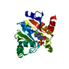
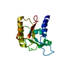
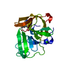
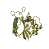
| Title | Crystal structure of reduced Ost6L | ||||||
 Components Components | Dolichyl-diphosphooligosaccharide-protein glycosyltransferase subunit OST6 | ||||||
 Keywords Keywords |  TRANSFERASE / OXIDOREDUCTASE / ACTIVE SITE LOOP / REDOX STATE / Membrane / Transmembrane TRANSFERASE / OXIDOREDUCTASE / ACTIVE SITE LOOP / REDOX STATE / Membrane / Transmembrane | ||||||
| Function / homology |  Function and homology information Function and homology informationMiscellaneous transport and binding events / oligosaccharyltransferase complex / protein N-linked glycosylation via asparagine / protein N-linked glycosylation / protein-disulfide reductase activity / Neutrophil degranulation / protein-containing complex assembly / endoplasmic reticulum membrane / endoplasmic reticulumSimilarity search - Function | ||||||
| Biological species |  Saccharomyces cerevisiae (brewer's yeast) Saccharomyces cerevisiae (brewer's yeast) | ||||||
| Method | X-RAY DIFFRACTION / FOURIER SYNTHESIS / Resolution: 1.96 Å | ||||||
 Authors Authors | Stirnimann, C.U. / Grimshaw, J.P.A. / Schulz, B.L. / Brozzo, M.S. / Fritsch, F. / Glockshuber, R. / Capitani, G. / Gruetter, M.G. / Aebi, M. | ||||||
 Citation Citation | Journal: Proc.Natl.Acad.Sci.USA / Year: 2009 Title: Oxidoreductase activity of oligosaccharyltransferase subunits Ost3p and Ost6p defines site-specific glycosylation efficiency. Authors: Schulz, B.L. / Stirnimann, C.U. / Grimshaw, J.P. / Brozzo, M.S. / Fritsch, F. / Mohorko, E. / Capitani, G. / Glockshuber, R. / Grutter, M.G. / Aebi, M. | ||||||
| History |
|
- Structure visualization
| Structure viewer | Molecule: MolmilJmol/JSmol |
|---|
- Downloads & links
-Download
| PDBx/mmCIF format | 3g9b.cif.gz | 45.7 KB | Display | PDBx/mmCIF format |
|---|---|---|---|---|
| PDB format | pdb3g9b.ent.gz | 31.9 KB | Display | PDB format |
| PDBx/mmJSON format | 3g9b.json.gz | Tree view | PDBx/mmJSON format | |
| Others |  Other downloads Other downloads |
-Validation report
| Arichive directory | https://data.pdbj.org/pub/pdb/validation_reports/g9/3g9bftp://data.pdbj.org/pub/pdb/validation_reports/g9/3g9b | HTTPS FTP |
|---|
-Related structure data


-Links
 PDBj
PDBj


- Assembly
| Deposited unit | 
| ||||||||
|---|---|---|---|---|---|---|---|---|---|
| 1 |
| ||||||||
| Unit cell |
|
-Components
| #1: Protein | Mass: 20342.777 Da / Num. of mol.: 1 / Fragment: N-terminal domain Source method: isolated from a genetically manipulated source Source: (gene. exp.) Saccharomyces cerevisiae (brewer's yeast)Gene: OST6, YML019W / Plasmid: pBAD-ost6L-His10 / Production host:  Escherichia coli (E. coli) / Strain (production host): Top 10 / References: UniProt: Q03723, EC: 2.4.1.119 Escherichia coli (E. coli) / Strain (production host): Top 10 / References: UniProt: Q03723, EC: 2.4.1.119 |
|---|---|
| #2: Water | ChemComp-HOH / Water Mass: 18.015 Da / Num. of mol.: 91 / Source method: isolated from a natural source / Formula: H2O Mass: 18.015 Da / Num. of mol.: 91 / Source method: isolated from a natural source / Formula: H2O |
-Experimental details
-Experiment
| Experiment | Method: X-RAY DIFFRACTION / Number of used crystals: 1 |
|---|
- Sample preparation
| Crystal | Density Matthews: 2.14 Å3/Da / Density % sol: 42.47 % |
|---|---|
| Crystal grow | Temperature: 277 K / Method: vapor diffusion, sitting drop Details: 18% PEG 3350, 50 mM Na-citrate, pH 3, protein solution buffer: 50 mM Tris-HCl, pH 8, mixing ratio 1.5 (protein):1 (precipitant), VAPOR DIFFUSION, SITTING DROP, temperature 277K |
-Data collection
| Diffraction | Mean temperature: 100 K |
|---|---|
| Diffraction source | Source: ROTATING ANODE / Type: ENRAF-NONIUS FR591 / Wavelength: 1.5418 Å |
| Detector | Type: MAR scanner 345 mm plate / Detector: IMAGE PLATE / Date: May 29, 2007 |
| Radiation | Monochromator: OSMIC MIRRORS / Protocol: SINGLE WAVELENGTH / Monochromatic (M) / Laue (L): M / Scattering type: x-ray |
| Radiation wavelength | Wavelength: 1.5418 Å / Relative weight: 1 |
| Reflection | Resolution: 1.96→30 Å / Num. obs: 12881 / % possible obs: 99.8 % / Observed criterion σ(I): -3 / Redundancy: 5.6 % / Biso Wilson estimate: 34.9 Å2 / Rsym value: 0.053 / Net I/σ(I): 20.3 |
| Reflection shell | Resolution: 1.96→2.06 Å / Redundancy: 5.6 % / Mean I/σ(I) obs: 3.4 / Rsym value: 0.559 / % possible all: 100 |
- Processing
| Software |
| |||||||||||||||||||||||||||||||||||||||||||||||||||||||||||||||
|---|---|---|---|---|---|---|---|---|---|---|---|---|---|---|---|---|---|---|---|---|---|---|---|---|---|---|---|---|---|---|---|---|---|---|---|---|---|---|---|---|---|---|---|---|---|---|---|---|---|---|---|---|---|---|---|---|---|---|---|---|---|---|---|---|
| Refinement | Method to determine structure: FOURIER SYNTHESIS Starting model: Ost6L structure solved by SAD (bromide signal) Resolution: 1.96→29.853 Å / SU ML: 0.36 / Isotropic thermal model: ISOTROPIC / σ(F): 1.42 / Phase error: 25.48 / Stereochemistry target values: ML
| |||||||||||||||||||||||||||||||||||||||||||||||||||||||||||||||
| Solvent computation | Shrinkage radii: 0.9 Å / VDW probe radii: 1.11 Å / Solvent model: FLAT BULK SOLVENT MODEL / Bsol: 38.514 Å2 / ksol: 0.403 e/Å3 | |||||||||||||||||||||||||||||||||||||||||||||||||||||||||||||||
| Displacement parameters | Biso mean: 36.1 Å2
| |||||||||||||||||||||||||||||||||||||||||||||||||||||||||||||||
| Refinement step | Cycle: LAST / Resolution: 1.96→29.853 Å
| |||||||||||||||||||||||||||||||||||||||||||||||||||||||||||||||
| Refine LS restraints |
| |||||||||||||||||||||||||||||||||||||||||||||||||||||||||||||||
| LS refinement shell |
|
