 Movie
Movie Controller


+ Open data

- Basic information
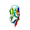
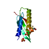
| Entry | Database: PDB / ID: 4qeg | ||||||
|---|---|---|---|---|---|---|---|
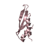
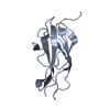
| Title | Crystal structure of domain I10 from titin (space group P41) | ||||||
 Components Components | Titin | ||||||
 Keywords Keywords | STRUCTURAL PROTEIN / Immunoglobulin domain / skeletal protein / sarcomere | ||||||
| Function / homology |  Function and homology information Function and homology informationsarcomerogenesis / structural molecule activity conferring elasticity / telethonin binding / skeletal muscle myosin thick filament assembly / cardiac myofibril assembly / muscle alpha-actinin binding / detection of muscle stretch / cardiac muscle tissue morphogenesis / regulation of catalytic activity / cardiac muscle hypertrophy ...sarcomerogenesis / structural molecule activity conferring elasticity / telethonin binding / skeletal muscle myosin thick filament assembly / cardiac myofibril assembly / muscle alpha-actinin binding / detection of muscle stretch / cardiac muscle tissue morphogenesis / regulation of catalytic activity / cardiac muscle hypertrophy / mitotic chromosome condensation / Striated Muscle Contraction / M band / actinin binding / I band / cardiac muscle cell development / regulation of protein kinase activity / sarcomere organization / structural constituent of muscle / skeletal muscle thin filament assembly / striated muscle thin filament / striated muscle contraction / protein kinase A signaling / cardiac muscle contraction / muscle contraction / condensed nuclear chromosome / positive regulation of protein secretion / Z disc / response to calcium ion / : / actin filament binding / Platelet degranulation / protein tyrosine kinase activity / protease binding / calmodulin binding / non-specific serine/threonine protein kinase / phosphorylation / protein serine kinase activity / protein serine/threonine kinase activity / calcium ion binding / positive regulation of gene expression / protein kinase binding / enzyme binding / extracellular exosome / extracellular region / ATP binding / identical protein binding / cytosolSimilarity search - Function | ||||||
| Biological species |  Homo sapiens (human) Homo sapiens (human) | ||||||
| Method | X-RAY DIFFRACTION / SYNCHROTRON / MOLECULAR REPLACEMENT / Resolution: 2 Å | ||||||
 Authors Authors | Bogomolovas, J. / Labeit, S. / Mayans, O. | ||||||
 Citation Citation | Journal: Open Biology / Year: 2016 Title: Exploration of the pathological potential of isolated mSNPs in titin: the cardiomyopathy-linked mutation T2580I Authors: Bogomolovas, J. / Labeit, S. / Anderson, B. / Williams, R. / Lange, S. / Simon, B. / Khan, M.M. / Rudolf, R. / Bullard, B. / Rigden, D.J. / Granzier, H. / Labiet, S. / Mayans, O. | ||||||
| History |
|
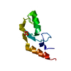
- Structure visualization
| Structure viewer | Molecule: MolmilJmol/JSmol |
|---|
- Downloads & links
-Download
| PDBx/mmCIF format | 4qeg.cif.gz | 51.7 KB | Display | PDBx/mmCIF format |
|---|---|---|---|---|
| PDB format | pdb4qeg.ent.gz | 36.9 KB | Display | PDB format |
| PDBx/mmJSON format | 4qeg.json.gz | Tree view | PDBx/mmJSON format | |
| Others |  Other downloads Other downloads |
-Validation report
| Arichive directory | https://data.pdbj.org/pub/pdb/validation_reports/qe/4qegftp://data.pdbj.org/pub/pdb/validation_reports/qe/4qeg | HTTPS FTP |
|---|
-Related structure data
| Related structure data | |
|---|---|
| Similar structure data |


-Links
 PDBj
PDBj


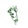
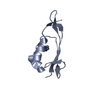
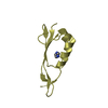
- Assembly
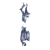
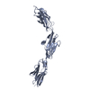
| Deposited unit | 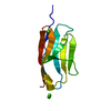
| ||||||||
|---|---|---|---|---|---|---|---|---|---|
| 1 |
| ||||||||
| Unit cell |
|
-Components
| #1: Protein | / Connectin / Rhabdomyosarcoma antigen MU-RMS-40.14 Mass: 10178.617 Da / Num. of mol.: 1 / Fragment: domain I10 Source method: isolated from a genetically manipulated source Source: (gene. exp.) Homo sapiens (human) / Gene: TTN / Plasmid: pETM11 / Production host:  Escherichia coli (E. coli) / Strain (production host): BL21 Escherichia coli (E. coli) / Strain (production host): BL21References: UniProt: Q8WZ42, non-specific serine/threonine protein kinase | ||
|---|---|---|---|
| #2: Chemical |   Mass: 40.078 Da / Num. of mol.: 3 / Source method: obtained synthetically / Formula: Ca Mass: 40.078 Da / Num. of mol.: 3 / Source method: obtained synthetically / Formula: Ca#3: Water | ChemComp-HOH / | Water Mass: 18.015 Da / Num. of mol.: 121 / Source method: isolated from a natural source / Formula: H2O Mass: 18.015 Da / Num. of mol.: 121 / Source method: isolated from a natural source / Formula: H2O |
-Experimental details
-Experiment
| Experiment | Method: X-RAY DIFFRACTION / Number of used crystals: 1 |
|---|
- Sample preparation
| Crystal | Density Matthews: 2.25 Å3/Da / Density % sol: 45.31 % |
|---|---|
| Crystal grow | Method: vapor diffusion, sitting drop / pH: 7.5 Details: 0.2 M CaCl2, 30% PEG 3350, 0.1 M Tris, 3% Isopropanol, pH 7.5, VAPOR DIFFUSION, SITTING DROP |
-Data collection
| Diffraction | Mean temperature: 100 K |
|---|---|
| Diffraction source | Source: SYNCHROTRON / Site: Diamond  / Beamline: I24 / Wavelength: 0.9464 Å / Beamline: I24 / Wavelength: 0.9464 Å |
| Detector | Type: DECTRIS PILATUS 6M / Detector: PIXEL |
| Radiation | Protocol: SINGLE WAVELENGTH / Monochromatic (M) / Laue (L): M / Scattering type: x-ray |
| Radiation wavelength | Wavelength: 0.9464 Å / Relative weight: 1 |
| Reflection | Resolution: 2→26 Å / Num. obs: 5665 / % possible obs: 93.2 % / Redundancy: 2.2 % / Rsym value: 0.139 / Net I/σ(I): 4.96 |
| Reflection shell | Resolution: 2→2.071 Å / Redundancy: 1.9 % / Mean I/σ(I) obs: 2.34 / Num. unique all: 560 / Rsym value: 0.3413 / % possible all: 91.9 |
- Processing
| Software |
| ||||||||||||||||||||||||||||||||||||||||
|---|---|---|---|---|---|---|---|---|---|---|---|---|---|---|---|---|---|---|---|---|---|---|---|---|---|---|---|---|---|---|---|---|---|---|---|---|---|---|---|---|---|
| Refinement | Method to determine structure: MOLECULAR REPLACEMENT / Resolution: 2→18.76 Å / SU ML: 0.26 / σ(F): 2.02 / Phase error: 24.89 / Stereochemistry target values: ML
| ||||||||||||||||||||||||||||||||||||||||
| Solvent computation | Shrinkage radii: 0.9 Å / VDW probe radii: 1.11 Å / Solvent model: FLAT BULK SOLVENT MODEL | ||||||||||||||||||||||||||||||||||||||||
| Refinement step | Cycle: LAST / Resolution: 2→18.76 Å
| ||||||||||||||||||||||||||||||||||||||||
| Refine LS restraints |
| ||||||||||||||||||||||||||||||||||||||||
| LS refinement shell |
| ||||||||||||||||||||||||||||||||||||||||
| Refinement TLS params. | Method: refined / Origin x: 21.2508 Å / Origin y: 21.273 Å / Origin z: 47.1884 Å
| ||||||||||||||||||||||||||||||||||||||||
| Refinement TLS group | Selection details: (chain 'A' and resid 2833 through 2921) |
